

Simultaneous AFM and Fluorescence Imaging of Living Cells
This publication presents a combined Atomic Force Microscopy (AFM) and epi-fluorescence microscopy setup for simultaneous imaging of live cells under physiological conditions. By integrating high-resolution AFM with fluorescence imaging, the method provides complementary structural and functional information about cellular processes.

The study demonstrates the system’s capabilities using Chinese Hamster Ovary (CHO) cells expressing eGFP-tagged scavenger receptor class B type I (SR-BI). Fluorescence imaging tracks labeled membrane components, while AFM resolves nanoscale surface structures. This dual approach enables detailed correlation between membrane receptor distributions and cytoskeletal features.
Fixed and live cells were imaged to illustrate the system’s flexibility. While fixed cells provided stable imaging conditions, live-cell imaging revealed the dynamic nature of soft membranes and underlying cytoskeletal structures. The fluorescence image aided in identifying cellular protrusions and lamellipodia, while AFM provided higher resolution of surface features.
This integration expands cellular imaging capabilities, enhancing understanding of structure-function relationships in live-cell environments.
Imaging DNA in Solution with the AFM
This publication explores the use of Atomic Force Microscopy (AFM) for imaging DNA molecules under physiological conditions with nanometer-scale resolution. DNA immobilization techniques on smooth mica substrates are emphasized as a prerequisite for stable imaging in liquid environments.
Various strategies are outlined to attach DNA to mica surfaces, including the use of divalent metal cations (e.g., Mg²⁺, Ni²⁺) for electrostatic bridging and chemical modifications with aminosilanes or ethanolamine to introduce positive charges. Among these, divalent cation-mediated attachment is highlighted for its simplicity and effectiveness in maintaining DNA stability during imaging.
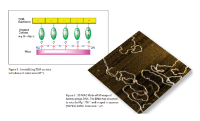
The study demonstrates high-resolution imaging of lambda phage DNA using MAC Mode AFM in aqueous buffer, providing three-dimensional views of the DNA structure. The approach enables visualization of supercoiled, looped, and kinked DNA under near-physiological conditions.
This work underscores the versatility of AFM as a tool for studying DNA and other biological molecules, offering insights into their structure and interactions at the nanoscale.
Imaging DNA with the 6000ILM AFM
This publication highlights the use of the Agilent 6000ILM Atomic Force Microscope (AFM) for imaging DNA molecules under both ambient and physiological conditions. The study emphasizes the importance of immobilizing DNA on smooth substrates like mica, which provides a negatively charged surface for electrostatic interactions with DNA’s phosphate backbone.

Using divalent cations such as Ni²⁺ and Mg²⁺, DNA was effectively immobilized on mica, facilitating high-resolution imaging in HEPES buffer. The study demonstrates the versatility of AFM for observing DNA conformations, substructures, and DNA-protein complexes in liquid environments. Imaging in physiological buffers allows for the investigation of DNA in states closely mimicking its native environment.
The results reveal that AFM is a powerful tool for understanding DNA’s role in gene expression, replication, and recombination. This approach offers insights into the nanomechanical properties and structural characteristics of DNA, advancing the study of nucleic acids in molecular biology and nanotechnology.
Attaching Antibodies to AFM Probes with the Sulfhydryl Reactive PEG Tether, NHS-PEG18-PDP
This publication outlines a protocol for attaching antibodies to Atomic Force Microscopy (AFM) probes using a sulfhydryl-reactive polyethylene glycol (PEG) tether, specifically NHS-PEG18-PDP. This technique enables the functionalization of AFM probes for single-molecule recognition studies, such as Molecular Recognition Force Microscopy (MRFM) and Topography and Recognition Imaging (TREC).
The process involves multiple steps, including surface cleaning, aminofunctionalization of AFM probes using alkoxy aminosilanes, PEGylation with NHS-PEG18-PDP, and antibody activation with sulfhydryl groups. The activated antibodies are then conjugated to the PEG-functionalized probes, forming a stable and selective biosensor.

The modified probes are optimized for investigating ligand-receptor interactions at the nanoscale, with applications in mapping chemical compositions and understanding molecular interactions, such as antibody-antigen binding. This method ensures high specificity and stability while maintaining probe functionality, providing a robust platform for advanced AFM-based biological and chemical research.
Using AFM to Characterize DNA Microarrays
This publication demonstrates the use of Atomic Force Microscopy (AFM) for characterizing DNA microarrays, focusing on structural uniformity and probe density at the nanoscale. DNA microarrays, composed of 60-mer oligonucleotide probes immobilized on silanized glass substrates, are critical for high-throughput gene expression analysis, genotyping, and other biological assays.

Using an Agilent 5500 AFM in Acoustic AC (AAC) mode, the study examined microarrays under ambient conditions. AFM imaging revealed distinct probe elements approximately 4.5 nm above the silanized glass substrate, with clumped yet uniformly distributed oligonucleotides. Surface roughness analysis quantified the contrast between probe regions (RMS value: 1.07 nm) and silanized glass (RMS value: 0.429 nm).
The findings highlight the advantages of AFM in detecting surface defects, ensuring quality control, and providing detailed topographic and structural information. This non-destructive method supports improved manufacturing and functional performance of DNA microarrays in research and clinical applications.
Diatoms: AFM Imaging of Natural Synthesis at the Nanoscale
This publication investigates the nanoscale structures of diatom silica skeletons using Atomic Force Microscopy (AFM). Diatoms, unicellular photosynthetic algae, synthesize intricate silica structures through energy-efficient, low-temperature processes. These natural systems offer valuable insights for nanotechnology, particularly in fabricating nanoscale devices.

The study focuses on two diatom types: centric and pennate. Centric diatoms, exemplified by Thalassiosira pseudonana, display radial symmetry with silica spokes composed of 20–30 nm nodules. Pennate diatoms, such as Navicula pelliculosa, exhibit bilateral symmetry with smooth silica ribs and ordered nanometer-scale pits. AFM imaging of these structures was performed on gelatin-coated mica, revealing their complex architectures.
Diatoms are ecologically significant, contributing to carbon dioxide fixation and oxygen production. Their reproducible nanoscale synthesis capabilities inspire biomimetic approaches in materials science. This study demonstrates AFM’s utility for high-resolution imaging of diatom skeletons, advancing the understanding of natural nanoscale fabrication processes.
MAC Mode AFM Studies of Zinc-Induced DNA Kinking
This publication investigates zinc-induced DNA kinking using MAC Mode Atomic Force Microscopy (AFM), emphasizing its high resolution and ability to image DNA under near-physiological conditions. DNA kinking, characterized by abrupt bending, is a critical structural change influencing gene expression, replication, and recombination. The study used 168-base-pair circular DNA molecules to analyze kinking behavior under varying Zn²⁺ concentrations.

At high Zn²⁺ concentrations (1 mM), DNA molecules exhibited multiple kinks regardless of Mg²⁺ levels, confirming Zn²⁺’s specific interaction with DNA bases. Reducing Zn²⁺ concentration to 100 µM still resulted in kinked structures, but at 50 µM, most DNA reverted to a circular form, identifying a threshold for zinc-induced kinking between 50–100 µM.

The cooperative nature of Zn²⁺-induced kinking and its impact on DNA structure were highlighted. The study also demonstrated improved AFM imaging contrast with reduced salt concentrations, underscoring MAC Mode AFM’s effectiveness for nanoscale DNA conformational studies.
The Evolution of Scanning Probe Microscopes for Biological Imaging
This publication reviews the evolution and applications of Scanning Probe Microscopy (SPM), particularly Atomic Force Microscopy (AFM), in biological research. Initially derived from Scanning Tunneling Microscopy (STM), AFM introduced a flexible cantilever system capable of imaging non-conductive and insulating materials, overcoming STM’s limitations. Since its commercial introduction in 1989, AFM has become integral to life sciences, enabling molecular and nanoscale imaging under physiological conditions.

The paper discusses AFM’s progression from contact mode to dynamic modes like Acoustic AC (AAC) and Magnetic AC (MAC), which allow for high-resolution imaging of delicate samples, such as DNA, proteins, and living cells, in liquid environments. MAC Mode is particularly highlighted for its gentle imaging capabilities, reducing sample deformation while providing clear resonance signals.
AFM’s applications now include DNA-protein interaction studies, force spectroscopy for molecular binding analysis, and integration with microarray technology, solidifying its role in modern biological and nanotechnology research.
Immobilizing Biological Molecules on AFM Probes for MRFM and TREC Studies
This publication details the immobilization of biological molecules on Atomic Force Microscopy (AFM) probes for advanced applications such as Molecular Recognition Force Microscopy (MRFM) and Topography and Recognition Imaging (TREC). The methodology involves attaching ligands (e.g., nucleic acids, antibodies) to AFM probes using polyethylene glycol (PEG) tethers. These tethers enhance molecular flexibility, allowing ligands to diffuse within defined volumes and reorient for optimal binding with substrate-bound receptors.

The document outlines key steps, including probe cleaning, amination, PEGylation, and ligand bioconjugation. PEG tethers, available in various lengths, optimize single-molecule resolution and reduce nonspecific interactions. For example, shorter PEG linkers (~8–10 nm) improve lateral resolution in TREC imaging. Advanced PEG linkers, such as NHS-PEG18-PDP, enable efficient ligand attachment, while thiolation processes prepare proteins for conjugation.
This immobilization strategy enhances AFM’s capabilities for studying biomolecular interactions at nanometer resolution, advancing applications in molecular biology, biophysics, and nanotechnology.
In Situ Studies of a Fungal Polysaccharide Using MAC Mode AFM
This publication investigates the in situ imaging of fungal polysaccharides using Magnetic AC (MAC) Mode Atomic Force Microscopy (AFM). The study focuses on scleroglucan, a (1→3)-linked β-D-glucan with (1→6) branching, which serves as a structural component in cell walls and exhibits immunostimulatory properties. Scleroglucan forms triple-helical structures that can be denatured into random coils and subsequently renatured into linear or cyclic configurations.

Using MAC Mode AFM, the study captures high-resolution images of renatured scleroglucan molecules deposited on mica in an aqueous environment. The imaging reveals distinct structural forms, with linear molecules exhibiting greater thickness than cyclic ones. The study also highlights MAC Mode’s ability to minimize sample damage and provide reproducible imaging of soft biological samples in liquid environments.
This work demonstrates MAC Mode AFM as a powerful tool for studying polysaccharide structures, offering insights into their molecular organization and potential applications in biological and material sciences.
Applications of MAC Mode AFM in Biology, Pharmaceutical, and Other Bio-Related Industries
This publication highlights the applications of Magnetic AC (MAC) Mode Atomic Force Microscopy (AFM) in biological, pharmaceutical, and cosmetic research. MAC Mode enhances AFM’s capabilities by minimizing sample damage, improving resolution, and enabling imaging under native or controlled conditions, such as buffered solutions or physiological temperatures.

The document demonstrates MAC Mode’s versatility through examples including structural biology, where it resolves DNA minicircles, nucleosomes, and bacterial S-layer proteins. MAC Mode provides insights into DNA behavior under different ionic conditions, such as kinking induced by Zn²⁺. It also resolves chromatin structures with nanoscale detail, aiding studies of epigenetic mechanisms.
In pharmaceutical research, MAC Mode images liposomes and drug carriers, such as lactose crystals, under varying environmental conditions. Similarly, in cosmetics, it visualizes changes in material surfaces, like human hair before and after treatment.
This publication underscores MAC Mode’s broad utility for non-invasive, high-resolution analysis across diverse fields, facilitating advancements in material and life sciences.
Scanning Probe Studies of a Metalloprotein
This publication explores the use of Scanning Probe Microscopy (SPM) methods, including Scanning Tunneling Microscopy (STM) and Atomic Force Microscopy (AFM), for studying metalloproteins under quasi-physiological conditions. It focuses on site-specific immobilization of cytochrome P450cam and azurin proteins on gold electrodes using engineered cysteine residues. This approach allows controlled orientation and high-resolution imaging of the proteins while preserving their activity.

High-resolution STM imaging reveals densely packed monolayers of cytochrome P450cam, showing molecular dimensions consistent with crystallographic data. Electrochemical studies confirm enhanced electron transfer between the immobilized enzyme and the electrode, attributed to the strategic positioning of the cysteine residue near the enzyme’s active site.
The study demonstrates the enzyme’s catalytic activity and electrochemical addressability, making this methodology valuable for biosensor development. The findings also highlight the potential for SPM techniques to resolve nanoscale structural and functional properties of metalloproteins in real-time.
In Vitro Studies of Microtubule Structures Using MAC Mode AFM
his publication explores the use of Magnetic AC (MAC) Mode Atomic Force Microscopy (AFM) for in vitro imaging of microtubule structures. Microtubules, composed of alternating α- and β-tubulin subunits, are dynamic cellular components involved in various biological processes, such as organelle positioning and chromosome segregation.

Using bovine brain tubulin polymerized with taxol, MAC Mode AFM enabled high-resolution imaging of microtubules under physiological buffer conditions. Individual tubulin subunits were resolved with dimensions of approximately 4 nm, consistent with electron microscopy data. Protofilaments and their interconnections were also visualized, with ridges separated by 4-nm-wide valleys.
The study highlights MAC Mode AFM’s capability to image soft biological structures with minimal sample deformation. Additionally, experiments with katanin, a microtubule-severing protein, demonstrated real-time disassembly of microtubules, revealing dynamic structural changes.
This work establishes MAC Mode AFM as a powerful tool for studying cytoskeletal components and their interactions under near-native conditions.
Topography and Recognition Imaging with PicoTREC, a Scanning Probe Microscopy Accessory for Mapping Target Molecules on a Sample Surface
This publication highlights the capabilities of PicoTREC, an advanced accessory for Atomic Force Microscopy (AFM), in topography and recognition imaging. By combining AFM with dynamic force microscopy (e.g., MAC Mode), PicoTREC enables simultaneous mapping of surface topography and specific molecular interactions at nanometer resolution.
The technology employs an AFM tip functionalized with a ligand tethered via a flexible polyethylene glycol (PEG) linker. This configuration allows the ligand to reorient for optimal binding with target molecules on the surface. During lateral scanning, PicoTREC detects binding events by monitoring changes in oscillation amplitude, creating a recognition map alongside the topographical image.

Applications include mapping antibody-antigen interactions and identifying receptor sites without the need for fluorescence or radioactive labels. For example, PicoTREC accurately visualized antihistone antibody binding to chromatin molecules. The system’s sensitivity to single-molecule interactions makes it a powerful tool for studying biomolecular recognition and advancing nanoscale biological research.
Simultaneous AFM/Fluorescence Imaging
of Living Cells – Fluorescence-guided Force Spectroscopy
This publication highlights the combined use of Atomic Force Microscopy (AFM) and fluorescence microscopy for high-resolution imaging and force spectroscopy of living cells. Using the Agilent 5500 ILM AFM/SPM system integrated with an inverted light microscope, the study demonstrates simultaneous visualization of cellular morphology and specific biomolecular interactions under physiological conditions.

The methodology involves Chinese hamster ovary (CHO) cells expressing eGFP-tagged scavenger receptor B type 1 (SRB1). Fluorescence microscopy identifies receptor distribution, while AFM captures nanoscale topographical details. Force spectroscopy with antibody-functionalized AFM tips quantifies receptor-ligand interactions, revealing unbinding forces of 64 and 85 pN for SRB1-antibody complexes.
This approach enables precise mapping of receptor sites, achieving a binding probability of 13.6% when guided by fluorescence imaging, compared to only 3-4% without guidance. The integration of AFM and fluorescence microscopy offers powerful insights into live-cell processes, advancing studies in molecular biology and receptor dynamics.
Molecular Recognition Imaging of Biological Systems with PicoTREC
This publication examines the capabilities of PicoTREC for molecular recognition imaging, combining topographic and recognition imaging to map biomolecular interactions with nanometer resolution. Using functionalized AFM tips with tethered molecules, PicoTREC detects specific binding events through force-based measurements, offering insights into receptor-ligand interactions.
The study demonstrates PicoTREC’s efficacy in various systems, such as avidin-biotin and anti-biotin IgG interactions on mica. Topographic images revealed molecular distribution, while corresponding recognition images confirmed specific binding sites.

For avidin-biotin, recognition signals aligned precisely with topographic features, showcasing high specificity. In anti-biotin IgG studies, individual antibody molecules were resolved, validating the approach for single-molecule analysis.
The technique was further applied to micropatterned biomolecules, distinguishing between regions of specific binding and non-specific adhesive forces. PicoTREC’s ability to analyze weak and strong interactions, combined with high spatial resolution, underscores its potential for investigating biomolecular affinity, orientation, and surface interactions in complex systems.