Atomic force microscopy (AFM) sidesteps that trade-off entirely. It's the only imaging technique that delivers sub-nanometer resolution on biological samples sitting in liquid, at room temperature, under conditions close to what a cell actually experiences.
This guide covers how AFM works, how researchers prepare biological samples for it, where it's being used in real biology labs, and what to look for when choosing a system.
Key Takeaways
- AFM images biomolecules and cells at nanometer-to-sub-nanometer resolution without staining or fixation
- Operates directly in liquid for live-cell and biomolecule imaging under near-physiological conditions
- Measures mechanical properties beyond topography: stiffness, elasticity, and adhesion
- Sample immobilization strategy makes or breaks a biological AFM experiment
- Purpose-built BioAFM systems with fluid stages and environmental control support live-cell and buffer-based experiments
What Is Atomic Force Microscopy in Biology?
Bio-AFM applies atomic force microscopy to biomolecules, cells, and tissues to study both their structure and their mechanics. Instead of relying on light or electrons, it uses a physical probe to feel the surface of a sample, point by point, building a 3D topographical map.
For biologists, that means visualizing proteins, DNA strands, lipid membranes, and even living cells while simultaneously measuring how stiff, sticky, or elastic they are.
Brief History of Bio-AFM
AFM has roots in scanning tunneling microscopy (STM), which Gerd Binnig and Heinrich Rohrer introduced in 1981 at IBM. Because STM required a conductive sample, it couldn't image most biological material directly.
Binnig, along with Christoph Gerber and Calvin Quate, solved that limitation with the original AFM paper published in Physical Review Letters in 1986. Commercial systems followed in the late 1980s. Since then, the field has developed gentler, more quantitative imaging modes specifically to handle delicate, easily damaged biological samples.
Why AFM Matters for Biology
Compared with electron microscopy (EM) or fluorescence imaging, AFM offers a distinct set of advantages:
- Requires no dehydration, staining, or vacuum
- Works in native buffers, cell-culture media, or even water
- Detects single-molecule interactions, such as antigen-antibody or ligand-receptor binding forces
- Preserves live cells well enough to watch them divide, move, or respond to stimuli
How Does Atomic Force Microscopy Work?
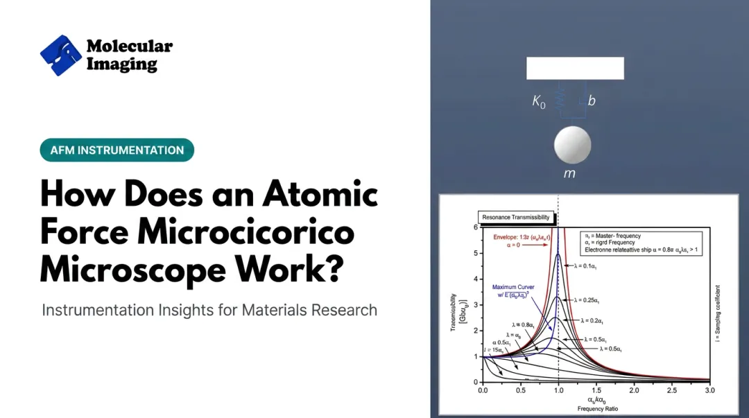
At its simplest, AFM uses a sharp probe mounted on a flexible cantilever. As the probe approaches a sample, forces such as Van der Waals interactions, electrostatics, and mechanical contact cause the cantilever to bend.
A laser beam reflects off the back of the cantilever onto a photodetector, which tracks this deflection with sub-nanometer precision. A piezoelectric scanner then moves the probe (or sample) across the surface in nanometer-scale steps, building an image line by line.

Imaging Modes That Matter for Biology
Three primary modes exist:
- Contact mode: the tip stays in constant contact with the sample, keeping force steady through feedback
- Tapping (intermittent contact) mode: the cantilever oscillates, touching the surface only briefly on each cycle
- Non-contact mode: the tip stays in the attractive force region above the surface, minimizing damage but more prone to interference from a water layer
Tapping mode is generally preferred for biological work because reduced lateral dragging protects fragile structures like cell membranes and microvilli.
Resolution You Can Expect
Published figures vary by sample and technique, so treat any single number cautiously. A 2024 biological-AFM review reports roughly 1 nm lateral resolution on isolated membrane patches and closer to 10 nm on living cells, since live cells move and deform during scanning. Vertical resolution is typically far better than lateral resolution in all cases.
Force Curves and Nanomechanical Mapping
Beyond topography, AFM records force-distance curves at each pixel (essentially a tiny push-and-release test). Techniques like PeakForce Tapping use these curves to map:
- Elastic modulus (stiffness)
- Adhesion
- Deformation and indentation
This is how researchers turn a simple image into a quantitative map of a cell's mechanical fingerprint.

How Do You Prepare Biological Samples for AFM?
Sample prep for biological AFM is minimal compared with EM, but immobilization still requires care.
Substrate Selection and Immobilization
Common substrates include mica, glass, and functionalized surfaces:
- Mica is cleaved to expose an atomically flat surface, then often treated with APS or APTES chemistry to bind DNA and proteins electrostatically
- Glass offers optical transparency for combined AFM and light-microscopy setups
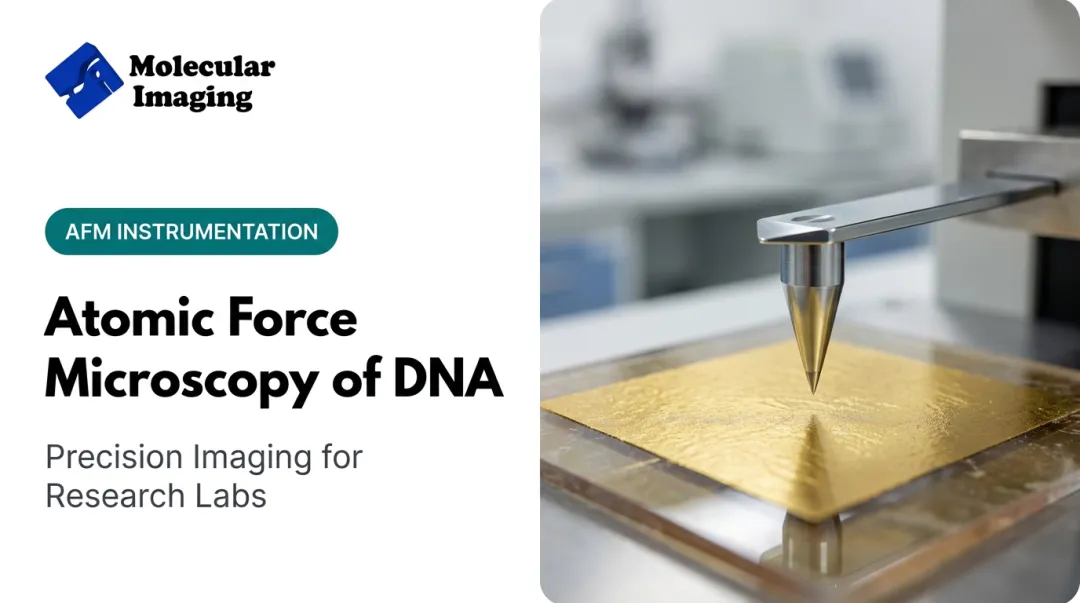
- Gold-coated substrates work well for thiol-based protein attachment
Live cells and bacteria need stronger adhesion strategies. A 2019 nanoscale bacterial imaging study found that Vectabond outperformed poly-L-lysine and gelatin across tested buffer conditions.
Molecular Imaging's mica substrates and gold-on-mica options give researchers a flat, reliable starting surface for DNA, proteins, and lipid bilayers.
Imaging in Liquid
To preserve native conformation, biological AFM is typically performed in buffer or cell culture media rather than air. This requires:
- A liquid cell or fluid chamber to contain the sample
- Cantilevers specifically rated for liquid operation (silicon or silicon-nitride tapping probes designed for aqueous environments)
- Stable temperature and pH control for extended live-cell sessions
Minimal processing is the real advantage here — no fixation, no heavy-metal staining, no conductive coating. Just a clean substrate, the right buffer, and a properly selected cantilever.
Key Applications of AFM in Biological Research
Imaging Biomolecules and Cellular Structures
Researchers use AFM to resolve biomolecular and cellular features at resolutions unreachable by light microscopy:
- DNA strands and individual proteins
- Lipid bilayer architecture
- Membrane surface features
Single-Molecule Force Spectroscopy
By functionalizing the AFM tip with a specific molecule, researchers can measure binding forces directly. One live-cell study using antibody-functionalized tips measured SR-BI receptor unbinding forces of 64 and 85 pN. Fluorescence guidance boosted binding probability to 13.6%, compared with just 3–4% without it.
Live-Cell Imaging
AFM captures dynamic biological processes as they happen:
- Cell division and membrane remodeling
- Cell adhesion and motility
- Real-time response to drugs or environmental changes
Disease and Diagnostic Research
Cell mechanics often shift with disease state. In a 2007 Nature Nanotechnology study, researchers measured stiffness in live metastatic cancer cells from patient pleural fluid and compared them with benign cells from the same samples.
Cross et al. found metastatic cancer cells were more than 70% softer than their benign counterparts, with a standard deviation more than five times narrower. That consistent mechanical signature is something optical imaging alone could not reveal.

AFM vs. Other Microscopy Techniques
| Feature | AFM | SEM | TEM | Confocal/Fluorescence |
|---|---|---|---|---|
| Sample prep | Minimal | Moderate, often coated | Extensive, thin-sectioned | Fluorescent labeling |
| Environment | Air, liquid, or vacuum | Usually vacuum | Vacuum | Air or liquid |
| Live-cell capable | Yes | No | No | Yes |
| Output | 3D topography + mechanics | 2D surface image | 2D internal structure | 2D/3D fluorescence |
| Lateral resolution | Sub-nm to a few nm | 1-10 nm | 0.1-0.2 nm | Diffraction-limited |
AFM's unique advantage is its combination of 3D topography and force data. Neither SEM nor TEM can offer that: both produce flat 2D images and require vacuum conditions incompatible with living samples.
Choosing the Right AFM System for Biological Research
Not every AFM setup is built for biology. Researchers working with live cells or delicate biomolecules need a system with real environmental control, not just a basic scanner.
Look for:
- Fluid stage compatibility for imaging directly in buffer or media
- Temperature control to maintain physiological conditions during extended sessions
- Low-drift design, since biological samples are often imaged over long periods
- Optical integration, useful for correlating AFM data with fluorescence
Molecular Imaging's 5500 Series, including the 5500 ILM PicoPlus BioAFM, was built with these needs in mind. It supports MAC Mode for gentle imaging of soft biological materials in fluids, liquid-cell designs, and a patented heating/cooling anti-drift stage rated from -30°C to 250°C. Environmental Isolation Chambers add control over humidity, gas composition, and low-oxygen conditions when needed.
For labs setting up their first bio-AFM workflow, Molecular Imaging also provides installation guidance, cantilever-alignment procedures, and a video-tutorial library covering sample prep and in-situ liquid imaging. Modular configuration support is available when a project calls for combining AFM with fluorescence or electrochemical measurements.
Frequently Asked Questions
What is the main purpose of atomic force microscopy in biology?
AFM's main purpose in biology is nanoscale visualization and mechanical characterization of biomolecules and live cells under near-physiological conditions. It reveals both structure and stiffness without staining or fixation.
How does atomic force microscopy work?
A sharp probe on a flexible cantilever detects surface forces as it scans a sample. A laser and photodetector track cantilever deflection, while a piezoelectric scanner moves the probe with nanometer precision to build an image.
How do you prepare biological samples for atomic force microscopy?
Samples are typically immobilized on mica or functionalized substrates, then imaged directly in buffer or liquid to preserve native structure. Processing is minimal compared with electron microscopy's fixation and coating requirements.
Can AFM image living cells?
Yes. AFM can image live cells directly in liquid without fixation, capturing dynamic processes like division, adhesion, and motility in near real-time.
What is the resolution of AFM compared to electron microscopy?
AFM offers comparable or superior vertical resolution to EM, with lateral resolution ranging from sub-nanometer to a few nanometers depending on the sample. It generally scans slower and covers a smaller field of view than EM.
What types of biological samples can be studied with AFM?
DNA, proteins, lipid membranes, viruses, bacteria, and live mammalian cells have all been successfully imaged with AFM, often in their native liquid environment.