Researchers studying outbreaks, developing diagnostics, or working on structural virology need something faster and less invasive than crystallography, and less prone to artifacts than standard electron microscopy. Atomic force microscopy (AFM) fills that gap. It images individual virus particles at nanometer resolution, in air or in liquid, without stains, dyes, or crystals.
This article covers how AFM detects and images viruses, how it stacks up against X-ray diffraction and electron microscopy, and what to look for in a BioAFM system if virus imaging is on your roadmap.
Key Takeaways
- AFM images intact, unlabeled viruses in near-native conditions, whether in air, liquid, or fixed on a substrate
- No crystallization or particle-averaging required, unlike X-ray diffraction or cryo-EM
- Tip shape and imaging mode control image accuracy, especially lateral measurements
- Documented structural virus studies include H1N1 influenza imaged by AFM
What Is Atomic Force Microscopy and Why Use It for Viruses
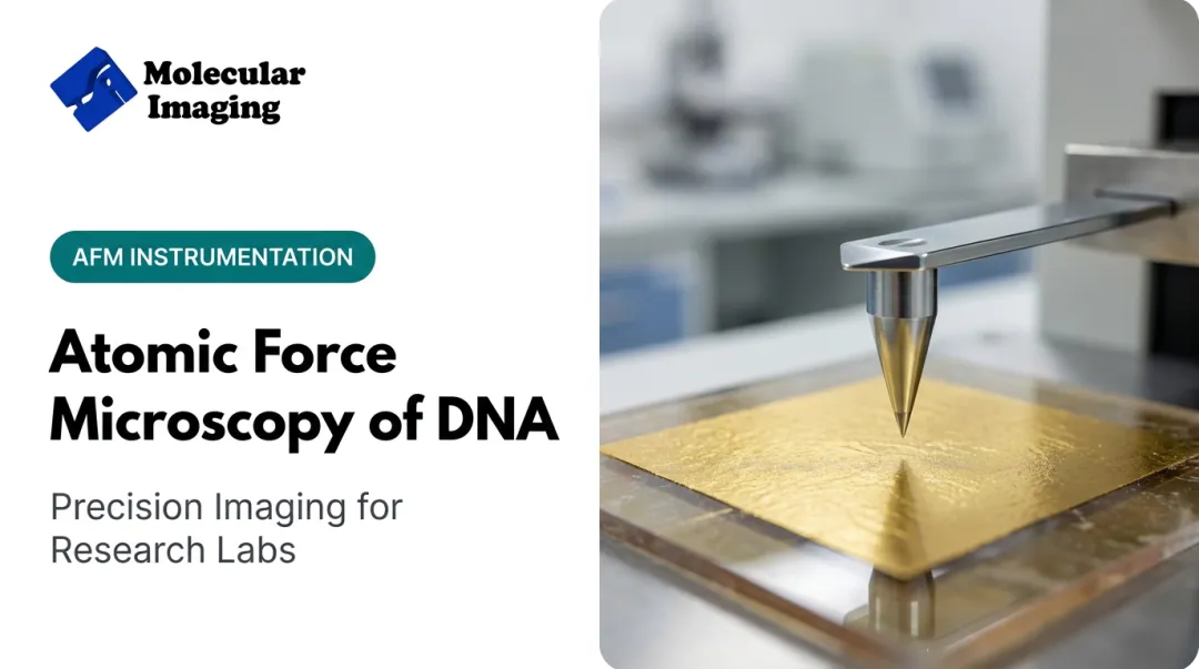
Atomic force microscopy (AFM) is a scanning probe technique. A cantilever with a nanoscale tip moves across a surface, sensing atomic-level forces and converting them into a topographical map. Instead of light or electrons, it "feels" the sample.
Why AFM Suits Virus Imaging
AFM can resolve capsid architecture, capsomere arrangement, and even DNA or RNA extruding from damaged particles. A 2011 virology review reports few-nanometer lateral resolution and roughly 0.5 nm height resolution, generating full three-dimensional topographical images of individual virions (Kuznetsov et al., 2011).
Key strengths:
- Label-free — no fluorescent tags, no heavy-metal staining
- Native conditions — imaging works in air, liquid, or on fixed samples
- In situ capability — viruses can be studied growing on infected cell surfaces
- Single-particle detail — no averaging across a population
Those same strengths show up in applied work. A peer-reviewed study used tapping-mode AFM to examine influenza A (H1N1) morphology, treating virions with Nonidet P-40 detergent to reveal internal structure as the envelope eroded (Hu et al., 2008). That study is a concrete case of AFM delivering structural data in influenza research.

How AFM Detects and Images Viruses: Core Mechanics
Contact Mode vs. Tapping Mode
In contact mode, the probe stays in near-constant contact with the surface as a piezoelectric stage moves it in a raster pattern. A laser bounces off the cantilever onto a photodiode, and converts those deflections into height data.
The problem: continuous contact creates lateral and shear forces. For a rigid material, that's fine. For a soft virus particle, it can flatten or displace the sample entirely.
Tapping mode (intermittent contact) solves this. The cantilever oscillates near its resonant frequency, touching the surface briefly instead of dragging across it. This is the preferred mode for biological samples:
- Minimizes friction and adhesion forces
- Reduces electrostatic disruption of soft particles
- Preserves capsid integrity during scanning
Substrate Preparation Matters
Viruses don't just sit still on glass or freshly cleaved mica. Most need a coating to immobilize them.
Poly-L-lysine is a common choice. A 2021 SARS-CoV-2 study coated mica with 0.1% poly-L-lysine for 10 minutes, then rinsed and deposited diluted virus directly into a liquid cell buffer (Lyonnais et al., 2021). Without the coating, the virus simply didn't stick to bare mica or glass.
A Real Detection Limit
Porter et al. (2006) captured feline calicivirus on an optimized substrate and used AFM as the imaging readout, reporting a detection limit of 3 x 10⁶ FCV/mL. That's a specific, quoted number from one workflow, not a universal AFM spec, but a useful benchmark for what's achievable with careful substrate design.
Beyond count-based detection, AFM's 3D topographic maps reveal features flat images miss: unusual mutant morphology, budding virions on infected cell surfaces, and growth patterns tracked over time on the same sample.
AFM vs. Other Virus Imaging Techniques
Every imaging technique makes a trade-off. Here's how the three major options compare for virus work:
| Criterion | AFM | X-ray Diffraction | TEM / Cryo-EM |
|---|---|---|---|
| Sample requirement | Native particle, no crystal needed | Requires a crystal, often 0.1 mm+ | Staining or vitrification required |
| What you see | Single-particle 3D surface | Averaged atomic structure | Averaged (cryo-EM) or stained (TEM) |
| Sample state | Air, liquid, or fixed | Crystallized, dry | Frozen or dehydrated |
| Prep complexity | Low to moderate | High (crystallization) | Moderate to high |
| Cost & footprint | Lower, benchtop-friendly | High | High |

X-ray diffraction delivers atomic-level detail, but only for viruses that crystallize well, which usually means small, symmetrical particles. Many viruses simply won't form usable crystals.
Cryo-EM and TEM bring their own limits. Negative-stain TEM can introduce drying and dehydration artifacts. Cryo-EM avoids staining but typically averages structure across thousands of particles to reach resolution, which can mask individual variation entirely.
AFM trades atomic resolution for simplicity: image one native particle, in a near-physiological state, without a synchrotron or a cryogenic chamber.
Interpreting AFM Virus Images: What Researchers Should Know
AFM images aren't a raw photograph of the virus. The tip itself is part of the measurement, and that matters for interpretation.
- Sharp tips give cleaner lateral resolution and less distortion
- Broad or dull tips make particles look wider than they are because the tip convolves with sample geometry
- Height data stays reliable — vertical measurements aren't affected by tip width the way lateral ones are
If your tip is worn or poorly matched to the sample scale, your reported virion diameter could be inflated well beyond the true value.

The fix: calibrate against known standards before drawing conclusions from lateral dimensions. A classic 1992 calibration study deduced tip shape by imaging biomolecules of known diameter, including DNA and tobacco mosaic virus (Thundat et al., 1992).
Grid or grating calibration samples serve a similar purpose today. Run one before a virus imaging session, not after.
Choosing the Right BioAFM System for Virus Research
Not every AFM setup handles biological samples well. For virus imaging specifically, a few features matter more than others:
- Tapping-mode capability — non-negotiable for soft, damage-prone particles
- Fluid-cell compatibility — needed for near-physiological imaging conditions
- Environmental control — temperature and humidity stability during long scans
- Modularity — the ability to switch configurations without buying a new system Molecular Imaging's 5500 ILM PicoPlus BioAFM was built around this kind of flexibility. It supports MAC Mode for soft biological materials in fluids, offers optical access from above, below, and the side of the sample, and works with a range of liquid-cell designs. Documented biological work on the platform includes DNA, proteins, chromatin, and cells in aqueous buffer, plus virus imaging in air on silicon substrates. For labs assembling a virus-imaging workflow, cantilever choice matters too. Molecular Imaging's catalog includes tapping-mode options built for soft samples:
- FM-10 Low Frequency Soft Tapping Cantilever — designed to minimize sample disruption
- Si-L-10 Liquid Tapping Cantilever — for biological samples imaged in aqueous conditions
- Si3N4-L-10 Liquid Tapping Cantilever — durable silicon-nitride option for extended liquid sessions Molecular Imaging provides BioAFM technical documentation, an "Immobilizing Bio Molecules" manual, and a 60-minute video lecture on in-situ liquid imaging. For labs with existing instruments, ZoomAFM covers calibration, diagnostics, and repair so tip condition and calibration stay consistent—both of which drive measurement accuracy on soft viral samples.

Frequently Asked Questions
Can you see viruses with atomic force microscopy?
Yes. AFM visualizes individual virus particles at nanometer resolution, including capsid structure and surface arrangement, without any staining or fluorescent labeling.
What is the main advantage of AFM over electron microscopy for virus imaging?
AFM avoids the fixation and staining artifacts common in TEM and doesn't require the vacuum conditions cryo-EM depends on. Samples can stay hydrated and close to their native state.
Can AFM image viruses in liquid environments?
Yes. Tapping-mode AFM paired with a fluid cell allows imaging of viruses in near-physiological liquid conditions, preserving structure that air-drying might distort.
Does AFM require special sample preparation for viruses?
Usually, yes. Substrates like mica often need a coating, such as poly-L-lysine, to immobilize virus particles so they stay stable during scanning.
How accurate are AFM measurements of virus size?
Height measurements are highly reliable. Lateral measurements can be skewed by tip shape and width, so calibration against known standards is important before trusting size data.
Is AFM used in real virus outbreak investigations?
Yes. AFM has contributed to structural studies of viruses tied to real outbreaks, including documented work examining influenza A (H1N1) morphology and internal structure.