You might also hear "cantilever probe" used in semiconductor wafer testing, referring to probe cards used to check chips before packaging. That's a completely different tool for a completely different industry. This article focuses on the AFM cantilever probe: what it's made of, how it works, the main types available, and how to pick the right one for your research.
Key Takeaways
- A cantilever probe is a microfabricated beam with a sharp tip that bends in response to nanoscale forces
- Spring constant and resonance frequency determine which imaging mode and sample type a probe suits best
- Contact, tapping, and specialized probes (bio, conductive, magnetic) serve distinct research needs
- Probe selection depends on sample hardness, desired resolution, and environment (air vs. liquid)
- Molecular Imaging's web store offers cantilever probes across all these categories for AFM/STM systems
What Is a Cantilever Probe?
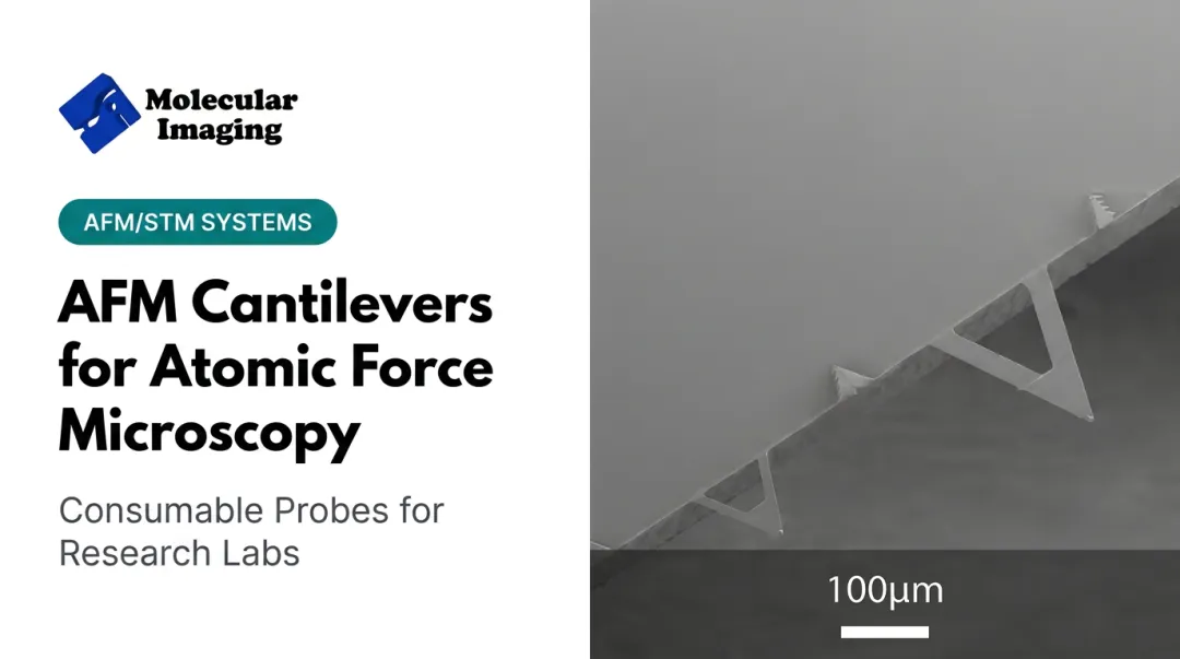
A cantilever probe is a microfabricated beam, typically silicon or silicon nitride, with an ultra-sharp tip mounted at one end and a fixed base at the other. Think of it as a diving board a few hundred micrometers long, with a needle-like point that can be just a few nanometers wide.
The physics is straightforward. As the tip approaches a sample surface, atomic, magnetic, or electrostatic forces cause the cantilever to flex or deflect. That deflection is the signal researchers use to build an image.
Key structural components include:
- Beam: flexible arm that bends under tip–sample forces
- Tip: nanometer-scale radius; sharper tips resolve finer detail
- Reflective coating: gold or aluminum layer used for optical detection
A laser bounces off the back of the cantilever onto a photodetector. As the cantilever bends, the reflected laser spot moves, and the photodetector converts that movement into a precise measurement of deflection.

Why Spring Constant and Resonance Matter
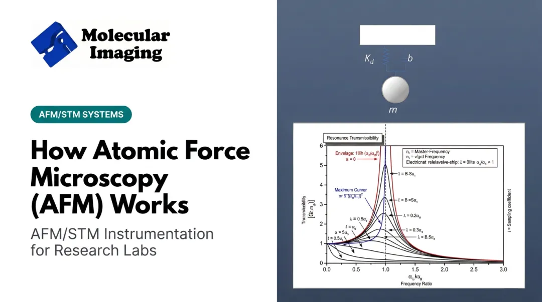
Two numbers define what a probe can do: spring constant (stiffness, measured in N/m) and resonance frequency (measured in kHz). According to catalog data from major AFM probe manufacturers, nominal spring constants across common products span roughly 0.07 to 42 N/m, with resonance frequencies from about 13 to 330 kHz, depending on the product line and intended imaging mode.
A soft, low-frequency probe suits delicate biological samples; a stiff, high-frequency probe suits hard materials like metals or ceramics. Matching those specs is easier when you can choose the right material and mode up front—Molecular Imaging's web store carries silicon and silicon nitride options, including the ContSi-10 contact-mode silicon cantilever and the S3N4-10 silicon nitride version, plus liquid-imaging variants for aqueous work.
How Cantilever Probes Work in AFM Imaging
Cantilever probes support three core imaging modes, each reading a different feedback signal from the cantilever.
Contact mode keeps the tip in continuous physical contact with the sample. The feedback loop monitors deflection directly and adjusts the sample height (via piezoelectric positioning) to maintain a constant force setpoint. This works well for rigid materials but can drag or damage soft samples.
Tapping/AC mode drives the cantilever to oscillate near its resonance frequency. The tip only touches the surface briefly during each cycle. As the tip-sample interaction changes, oscillation amplitude shifts, and the feedback loop adjusts height to hold that amplitude steady. That brief contact cuts lateral drag.
Non-contact mode keeps the tip hovering just above the surface, sensing attractive forces only. The cantilever's effective resonance frequency shifts slightly as attraction changes, and that shift becomes the feedback signal.

Matching Stiffness to the Sample
Probe sharpness and cantilever stiffness together determine what you'll actually see:
- Tip radius dominates spatial resolution : sharper tips resolve smaller features
- Spring constant and resonance frequency control force sensitivity and how much bandwidth you have for scanning
- System noise and sample compliance also affect final image quality, not stiffness alone
Bruker's own FAQ documentation notes that resolution depends on tip condition, system noise, and the sample itself — not spring constant in isolation. There's no single "stiffer is better" rule—match the probe to the job.
Types of Cantilever Probes
Cantilever probes are grouped by imaging mode and application. Matching the probe to your sample and measurement goal is the first step toward reliable AFM data.
Contact Mode Probes
Contact mode probes typically use lower spring constants, often well under 1 N/m, allowing continuous tip-sample contact without excessive force. They're common in materials characterization where the sample can tolerate direct contact.
Molecular Imaging's lineup includes:
- ContSi-10 — silicon, optimized for high-resolution topography
- S3N4-10 — silicon nitride, built for durability in direct-contact imaging
- Conductive Contact Cantilever — enables simultaneous topographical and electrical measurement
Tapping/AC Mode Probes
Tapping mode probes are stiffer than contact-mode levers and run at higher resonance frequencies. That lets the cantilever oscillate near the surface without dragging across it, which protects fragile samples and speeds up scanning.
Molecular Imaging's tapping-mode lineup includes:
- NCH-30-10 — high frequency, fast imaging in air
- NCL-30-10 — mid frequency, balanced resolution and stability
- FM-10 — low frequency, gentle enough for delicate biological or soft materials
Specialized Probes: Bio-AFM, Conductive, Magnetic
Beyond the standard modes, specialty probes cover liquid, electrical, magnetic, and mechanical measurements:
- Liquid tapping cantilevers (Si-L-10, Si3N4-L-10) for imaging biomolecules and cells in aqueous buffer
- MAC Mode levers (Types I, II, V, VI, VII) with paramagnetic coatings, tuned for anything from stiffer samples to soft targets like DNA and proteins
- EFM-10 for surface potential and electrical property mapping
- MFM-10 for magnetic domain imaging
- Diamond-10 diamond-coated tips for indentation and wear-resistant mechanical testing
These options support BioAFM and electrochemistry work, where standard air-imaging probes won't hold up.

Applications of Cantilever Probes in Research
Materials science. Contact-mode probes with compliant cantilevers around 0.1 N/m track adhesion-force changes in polystyrene films 18 to 127 nm thick. A 2018 AFM study documented shifts in glass-transition temperature as film thickness decreased.
This kind of force-distance measurement, not just imaging, is where thin-film and polymer researchers get their real data.
Life sciences. Liquid-imaging cantilevers let researchers visualize living cells without drying them out. One study used a probe with a spring constant of just 0.0611 N/m to resolve individual microvilli on living MDCK cells.
Those features measured roughly 1 micrometer in length and 200 nanometers in diameter, at peak forces as low as 100–130 pN. Molecular Imaging's Si-L-10 and Si3N4-L-10 cantilevers, paired with the 5500 ILM PicoPlus BioAFM system, target exactly this kind of soft, wet imaging.
Battery and electrochemistry research. In-situ electrochemical AFM studies use contact-mode silicon nitride cantilevers inside controlled environments such as argon glove boxes. Researchers watch electrode surfaces change in real time as voltage is applied.
Molecular Imaging supports this work through its Conductive Contact Cantilever, liquid-tapping probes, and PT-10 etched platinum-iridium STM tips, all relevant to labs working on magnesium-ion or lithium-ion electrode surfaces.

How to Choose the Right Cantilever Probe
Choosing a probe comes down to three questions:
- What's your sample? Hard materials tolerate stiffer, higher-force probes. Soft or biological samples need low spring constants and gentle contact.
- What imaging mode fits your goal? Contact mode for direct force measurement; tapping for speed with less sample damage; non-contact for the most delicate specimens.
- What resolution do you need? Smaller tip radius generally means finer detail, but only if system stability and sample condition cooperate.
Mismatched probes cause real problems:
- Too stiff on a soft sample → sample damage or deformation
- Too soft on a hard, rough sample → poor tracking, noisy images
- Wrong tip radius → blurred or misleading topography
Molecular Imaging supports this choice with practical resources:
- AFM Cantilevers Catalog
- "How to Choose Your MAC Lever" guide
- Cantilever mounting instructions
- Video tutorials on contact mode, tapping mode, EFM, KFM, and MFM
If you're still unsure, reach the ZoomAFM support team at 1-888-644-0389 or songxu@nanocuetech.com.
Frequently Asked Questions
What is a cantilever probe used for?
It's the sensing element in AFM. Researchers use it to image surface topography and measure atomic, magnetic, or electrostatic forces at the nanoscale in materials science, biology, and electrochemistry.
What is wafer probing and what is it used for?
Wafer probing is a semiconductor testing process, unrelated to AFM. It uses probe cards to test a chip's electrical performance while it's still on the wafer, before packaging.
What is a probe card used for?
A probe card is an electromechanical interface with multiple pins that contact chip pads during wafer testing. It belongs to the semiconductor industry, not AFM research.
How sensitive is an AFM cantilever probe?
Sensitivity varies by mode and setup. Some systems achieve force control down to 10 pN in air and fluid, while other imaging protocols operate around 100–130 pN. It depends heavily on the specific probe and instrument.
Can cantilever probes be reused or replaced?
Probes wear over time, especially the tip, and eventually need replacement rather than reuse. Molecular Imaging's web store carries a full range of replacement cantilevers, from contact and tapping probes to specialized diamond-coated and liquid-imaging options.