Many researchers struggle to measure these forces accurately. Traditional biology tools weren't built for nanoscale mechanics. You need something that can apply and measure piconewton-scale forces while imaging live cells in their natural liquid environment. That's where atomic force microscopy (AFM) comes in.
This guide covers how AFM enables mechanobiology research, the core techniques driving discoveries, real applications across disease research, and what to look for when selecting a BioAFM system.
Key Takeaways
- AFM resolves forces as small as 10⁻¹¹ N on live cells and tissues—no fixation required
- Force spectroscopy and nanoindentation link stiffness shifts to cancer, fibrosis, and regeneration
- Temperature control and liquid perfusion keep data physiological and reproducible
- Liquid-compatible, low-drift BioAFM hardware matters as much as the method itself
What Is Mechanobiology and Why It Matters
Mechanobiology studies how physical forces and biochemical signals jointly shape cell and tissue behavior. Cells don't just sit passively in their environment. They sense stiffness, tension, and geometry, then translate those mechanical cues into biological responses like gene expression changes or shifts in cell shape.
This isn't a new idea. D'Arcy Wentworth Thompson proposed as early as 1917, in On Growth and Form, that biological growth follows physical and mathematical principles. His hypothesis waited nearly a century for the imaging tools to validate it at the cellular scale.
Research areas shaped by mechanobiology include:
- Cancer metastasis and tumor progression
- Fibrosis and tissue scarring
- Wound healing and skin regeneration
- Cardiovascular disease and vascular remodeling
- Tissue engineering and regenerative medicine
Research on skin wound healing confirms that mechanical force significantly affects regeneration outcomes and acts as a primary driver of how tissue rebuilds itself.
How Atomic Force Microscopy Powers Mechanobiology Research
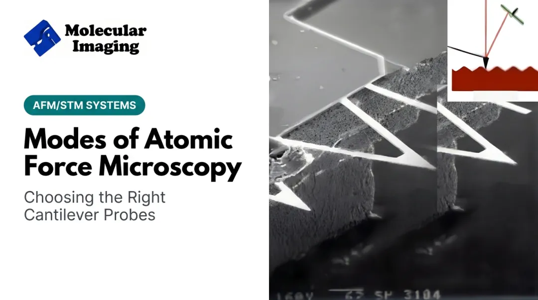
AFM works by scanning a nanometer-scale tip attached to a flexible cantilever across a sample. Tip-sample interactions bend the cantilever, and a laser reflected into a photodiode converts that deflection into a precise force measurement using Hooke's law.

This gives researchers something optical microscopy can't: direct mechanical data, not just visual images.
Why Liquid Compatibility Matters
Unlike vacuum-based imaging methods, AFM operates in physiological buffer. Cells stay alive, unfixed, and functional throughout the experiment. You can watch a cell respond to a mechanical stimulus in real time, in conditions that resemble its native environment.
Core Measurement Techniques
- Force spectroscopy: indents the cell membrane and records force curves to measure stiffness, elasticity, and adhesion
- Nanoindentation: quantifies Young's modulus of cells, extracellular matrix, and tissue samples
- Dynamic time-lapse imaging: captures real-time cellular responses to chemical or mechanical stimuli
The Cancer Stiffness Signature
A 2016 study using AFM on ovarian cells reported clear Young's modulus differences across cell lines:
- Healthy IOSE ovarian epithelial cells: 2.472 kPa
- HEY ovarian cancer cells: 0.884 kPa
- More aggressive HEY A8 line: 0.494 kPa
Softer cells tracked with more aggressive cancer. That stiffness gradient is a quantitative signal only force-resolved tools like AFM can capture.

Core AFM-Based Techniques Used in Mechanobiology
Several AFM techniques form the backbone of mechanobiology experiments, from single-molecule pulls to live-cell mapping under controlled conditions.
Single-Molecule and Single-Cell Force Spectroscopy
By attaching a cantilever to individual molecules, researchers can pull on receptor-ligand pairs and watch proteins unfold in real time. This reveals binding strength, folding kinetics, and membrane mechanics at a resolution few other tools can match.
Substrate Stiffness Mapping
Matrix rigidity directs stem cell fate. A landmark 2006 Cell study showed soft, brain-like substrates pushed mesenchymal stem cells toward neurogenic paths. Stiffer, muscle-like matrices (roughly 8-17 kPa) triggered myogenic differentiation, while rigid, bone-like surfaces drove osteogenic outcomes.
Multiparametric Imaging
Modern AFM modes collect topography and mechanical data simultaneously: stiffness, adhesion, and deformation in a single scan pass. You get structural detail and force data from the same scan, which matters when samples change quickly or cannot be re-imaged.
Environmental Control for Live-Cell Measurements
Live-cell mechanics data is only meaningful if the cell stays alive and stable during measurement. That requires:
- Temperature-controlled stages to maintain physiological conditions
- Environmental isolation chambers for humidity, gas, and low-oxygen control
- Anti-drift design to prevent measurement artifacts during long experiments
Molecular Imaging's temperature-controlled sample stage supports −30°C to 250°C with a patented anti-drift design. Its environmental isolation chamber manages humidity, temperature, low-oxygen conditions, and gas environments. Together, both keep live samples stable during extended force measurements.

Correlative Fluorescence Approaches
Combining AFM with fluorescence microscopy adds molecular identity to mechanical data. In one documented application, fluorescence-guided force spectroscopy on living CHO cells achieved a 13.6% binding probability, compared with just 3-4% without fluorescence guidance. The same work measured unbinding forces of 64 and 85 pN using antibody-functionalized tips.
Applications Across Research Fields
AFM mechanobiology shows up wherever cell or tissue mechanics drive function. Across labs, the same force-sensing toolkit answers different biological questions:
| Field | What AFM Reveals |
|---|---|
| Cancer research | Stiffness shifts tied to metastatic potential; a 2025 review links ductal stiffening in benign disease with softer phenotypes in invasive cells |
| Regenerative medicine | Scaffold roughness and mechanics that predict stem-cell adhesion and differentiation |
| Cardiovascular research | Direct cardiomyocyte contractility—e.g., forces rising from 1.74 to 3.49 nN as cells mature |
| Neuroscience | Cortical neuron elasticity mapped at 0.4–2.3 kPa under varying substrate stress |
| Infectious disease | Pathogen–host adhesion, including ~500 nN Staphylococcus aureus attachment to skin cells |

What unites these fields is a need to quantify force and mechanical phenotype when chemistry alone cannot explain the biology.
Choosing the Right AFM System for Mechanobiology Research
Not every AFM platform is built for live-cell work. Mechanobiology demands specific capabilities that general-purpose systems may lack.
Essential Capabilities for Live-Cell Work
Prioritize these features when you evaluate a platform:
- Liquid-imaging stability: Live cells need low-drift systems. Molecular Imaging's 5500 ILM PicoPlus BioAFM uses a lightweight, rigid mechanical loop and a pendulum multipurpose scanner to minimize drift during extended liquid imaging.
- MAC Mode compatibility: Magnetic AC mode supports gentle imaging of soft biological materials without damaging delicate structures.
- Optical integration: The 5500 ILM platform offers optical access from above, the side, and below the sample for correlative fluorescence work.
- Environmental accessories: Temperature-controlled stages and isolation chambers maintain physiologically relevant conditions during long experiments.
- Proper cantilevers: Liquid-tapping cantilevers such as the Si-L-10 and Si3N4-L-10 suit biological samples; the Diamond-10 supports indentation and mechanical testing.
Long-Term Reliability Matters Too
Mechanobiology programs run for years, not weeks. Calibration drifts, probes wear out, and systems need servicing.
ZoomAFM, Molecular Imaging's dedicated support service, provides hardware and software diagnostics, preventive maintenance, and precision calibration. It keeps existing AFM systems, including legacy Agilent 5500 platforms, running accurately.
Calibration samples such as the 500nm grid and grating standards help verify scanner accuracy before force experiments. Confirm a solid topographic baseline before you start measuring mechanics.
Frequently Asked Questions
What is the difference between AFM and traditional microscopy in mechanobiology?
AFM measures physical force and topography at nanoscale resolution by physically scanning a cantilever tip across the sample. Optical microscopy only provides visual images and can't directly quantify mechanical properties like stiffness or adhesion.
How does AFM measure the stiffness of a living cell?
A cantilever tip indents the cell membrane while recording force-versus-indentation curves. Researchers fit these curves using models like Hertz or Sneddon to calculate the cell's apparent Young's modulus.
Can AFM be used on live cells and tissues?
Yes. AFM operates directly in physiological buffers and liquid environments, often with temperature control, so you can image live cells and tissues without fixation.
What are common applications of AFM-based mechanobiology in disease research?
AFM is widely used in cancer research (stiffness as a metastasis marker), fibrosis studies, cardiovascular disease (cardiomyocyte contractility), and regenerative medicine (scaffold and stem cell mechanics).
What equipment is needed to start AFM-based mechanobiology experiments?
You'll need a BioAFM system capable of liquid imaging, environmental control accessories like temperature stages, liquid-compatible cantilevers, and calibration samples to verify measurement accuracy.
How do researchers maintain and service AFM systems over time?
Routine calibration, probe replacement, and professional diagnostic support keep systems accurate over years of use. Services like ZoomAFM provide remote diagnostics and preventive maintenance for existing AFM/STM platforms.