Introduction to AFM Contact Mode
Every researcher running an atomic force microscope faces the same trade-off: push for speed, sacrifice resolution. Push for gentleness, sacrifice throughput. Nanoscale topography doesn't come free, and the mode you choose determines what you give up.
Contact mode AFM is the most direct answer to that trade-off. It's the original scanning technique, where the tip stays in continuous contact with the sample surface while the system builds an image line by line.
This guide breaks down how contact mode actually works, its major configurations, when it beats tapping or non-contact mode, and what to look for in an AFM system built to run it well.
Key Takeaways
- Contact mode drags the tip across the surface, using cantilever deflection as feedback
- Works best on hard, rough, or robust samples where speed matters more than gentleness
- Lateral force mode, a contact mode variant, maps friction and material composition
- Force control below ~100 pN makes contact mode viable on delicate biological samples in liquid
- Modularity and cantilever choice determine how well a platform handles contact mode
What Is AFM Contact Mode?
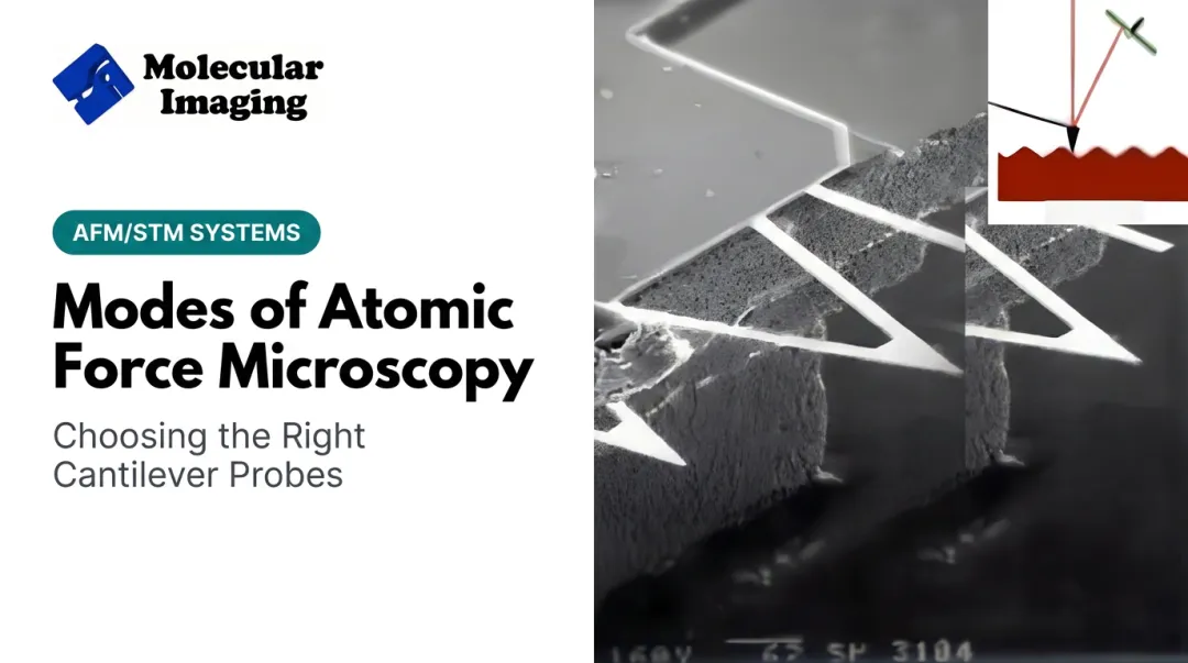
Contact mode is a static, non-oscillating imaging technique. Unlike tapping or non-contact modes, the cantilever isn't driven near its resonance frequency. Instead, the tip physically touches the sample and gets dragged across it during raster scanning.
The physics behind this is simple. As the tip meets a bump, dip, or edge, the cantilever bends. This bending follows Hooke's Law:
F = -kx
Here, k is the cantilever's spring constant and x is how far it deflects. A laser bounces off the back of the cantilever onto a photodetector, and that reflection tracks deflection in real time.
How the Feedback Loop Keeps Things Stable
An electronic feedback loop watches this deflection signal continuously. It adjusts the probe's z-height to hold deflection at a target value, called the deflection setpoint. When the tip hits a taller feature, the loop pulls the scanner back. When it hits a valley, the loop pushes forward.
This constant adjustment does two things:
- Keeps tip-sample force roughly constant across the scan
- Generates the z-height data that becomes your topography image
Snap-In and the Repulsive Force Regime
As the tip approaches the surface, attractive forces (van der Waals, capillary) build up. Once the force gradient exceeds the cantilever's stiffness, the tip suddenly jumps into contact. This is called snap-in, and it's an instability, not a gradual transition.
Once in contact, imaging happens on the repulsive branch of the force curve. This is what makes contact mode stable: the tip pushes back against the sample rather than hovering in an unstable attractive zone.
That repulsive contact still needs careful force control. Low spring-constant cantilevers matter here: a soft cantilever deflects enough for a clean signal while keeping force on the sample low enough to avoid damage.

Contact mode was the first AFM operating mode, introduced by Binnig, Quate, and Gerber in their original 1986 Physical Review Letters paper. Understanding it remains the foundation for every other AFM technique.
How Contact Mode AFM Operates: Key Configurations
Contact mode isn't a single fixed setup. Depending on what the feedback loop holds constant, you get several distinct configurations, each suited to different samples and goals.
Constant Force (Deflection) Mode
This is the standard setup most researchers default to. You set a target deflection, and the feedback loop works continuously to maintain it. The output is:
- A height (topography) channel, from the z-motion needed to hold deflection constant
- A deflection/error channel, showing how well the loop tracked the setpoint
Scan speed here is limited by how fast the z-feedback loop can respond to changing topography.
Constant Height Mode
Here, the probe's z-height stays fixed. No active force feedback runs during the scan. Instead, cantilever deflection itself becomes the image signal.
This only works on very flat, rigid samples, since large surface relief can generate excessive force or knock the tip out of contact entirely. It's used mainly for atomic-resolution imaging, where feedback bandwidth would otherwise limit resolution.
Error Signal (Error Mode) Imaging
During constant-force scanning, the residual deflection (the "error" the loop couldn't fully correct) gets captured as its own channel. This error signal often reveals fine edges and subtle surface details that the finite-bandwidth feedback loop smoothed over in the height image. Think of it as a sharpening pass on your topography data.
Lateral Force (Friction) Mode
Lateral force microscopy (LFM) measures a different kind of cantilever motion: torsion, caused by the tip twisting as it scans perpendicular to its long axis. This torsion maps frictional and material contrast across the surface.
Standard practice for isolating true friction signal:
- Scan the same line in the forward direction, then the reverse direction
- Compare signals: frictional torsion flips sign with scan direction, while topographic slope crosstalk doesn't
- Subtract the forward and reverse lateral signals to cancel out topographic crosstalk and isolate friction contrast

Advantages and Disadvantages of Contact Mode
Contact mode remains useful for specific jobs, but it comes with real limitations.
Advantages:
- High scan speeds on suitable samples, since there's no oscillation cycle to manage
- Strong topographic tracking on samples with large or abrupt height changes
- Simpler electronics and lower system cost compared to dynamic modes like tapping
Disadvantages:
- Continuous sliding generates lateral (shear) forces that can distort or displace soft, weakly attached samples
- In ambient air, capillary forces from adsorbed water layers add unwanted adhesion between tip and sample
- Tip wear and frictional artifacts can alter what looks like topography
The Biological Exception
Contact mode's reputation for damaging soft samples isn't the whole story. When interaction forces are tightly controlled, typically kept below roughly 100 pN, contact mode can image delicate biological structures successfully, especially in liquid where the ambient capillary meniscus disappears entirely.
A well-documented example: researchers used contact-mode high-speed AFM to image native purple membrane containing bacteriorhodopsin trimers in a Tris-HCl/KCl buffer (Biophysical Journal).
At an applied load of about 50 pN, they resolved individual protein molecules with lateral resolution near 20 angstroms, and the lattice stayed intact through long imaging sessions. That result challenges the "contact mode destroys soft samples" assumption, but only with disciplined force control.
Contact Mode vs. Tapping Mode vs. Non-Contact Mode
The three primary AFM operating modes split along one main axis: whether, and how, the cantilever touches the sample.
- Contact mode keeps the tip in continuous contact. No oscillation.
- Tapping mode oscillates the cantilever near resonance, touching the surface only briefly on each cycle.
- Non-contact mode also oscillates, but stays close enough to sense long-range attractive forces without deliberately touching the surface.
That difference in contact behavior drives everything else: force levels, damage risk, and where each mode gets used.
| Factor | Contact Mode | Tapping Mode | Non-Contact Mode |
|---|---|---|---|
| Lateral force | Highest (continuous drag) | Greatly reduced (brief contact) | Minimal |
| Damage risk | Highest for soft/mobile samples | Lower, preferred for fragile samples | Lowest under stable operation |
| Resolution | Atomic-level possible on stable samples | High for general-purpose imaging | Very high on clean surfaces |
| Typical environment | Air or liquid, hard/stable samples | Most common ambient mode; liquid also possible | Best in vacuum or controlled conditions |
Why Non-Contact Struggles in Ambient Air
A common misconception is that non-contact mode should work anywhere tapping mode does. It doesn't, and the reason is water. In ambient conditions, a thin adsorbed water film sits on both tip and sample. When they get close, this film forms a capillary bridge that pulls the tip into contact, defeating the whole point of "non-contact" operation.
That instability is exactly why tapping mode became the dominant ambient-condition alternative to contact mode. Its brief, repeated contact cycles punch through the water layer without letting sustained lateral drag build up.

Applications of Contact Mode AFM
Contact mode works best on samples that can tolerate direct, sustained tip contact, and where speed or friction data matter more than gentleness.
Hard, robust surfaces
- Inorganic crystals and semiconductor surfaces
- Thin films
- Rough industrial materials where fast scanning matters more than minimizing force
Material composition mapping via lateral force mode
LFM's real value shows up when height data alone can't distinguish surface components. In one well-cited study, researchers used LFM to map surface-phase distribution in a ternary polymer blend, resolving how the components intercalated at both micrometer and sub-100-nanometer scales (Surface Science, Cyganik et al.).
That friction contrast is often invisible in a standard height channel.
Biological niche applications
Membrane protein arrays imaged in liquid remain one of contact mode's most demanding—and successful—use cases. With carefully controlled forces, native structural detail stays intact instead of being lost under excessive load.
Choosing the Right AFM System for Contact Mode Imaging
Getting good contact mode results depends less on the mode itself and more on the hardware supporting it. Three things matter most:
- System modularity, so you can switch between contact, tapping, and other modes without buying separate instruments
- Sensitive deflection detection electronics, since contact mode lives and dies on clean deflection signal
- Low-spring-constant cantilevers matched to your sample type
Our AFM/STM platforms at Molecular Imaging are built around this kind of flexibility. The 5500 PicoPlus AFM+STM and 5420 PicoAFM both use a modular nose cone architecture, so researchers can swap between contact mode, tapping mode, and other techniques on the same base system.
Both offer dual scan ranges (roughly 9 µm to 90 µm) and support a wide range of liquid cell configurations. That setup suits contact mode in fluid—for example, silicon atomic-step imaging on the 5500 using ethanol as the medium.
For biologically focused work, the 5500 ILM PicoPlus BioAFM shares the same scanner architecture with optical access from above, below, and the side—so you can watch the sample while running contact mode in liquid or air.
On the cantilever side, we carry both silicon (ContSi-10) and silicon nitride (S3N4-10) contact mode probes, suited to different sample hardness and durability needs.

If you already own a contact-mode-capable system that's aging or acting up, ZoomAFM repair and service may be enough. It covers diagnostics, calibration, and remote or on-site support—often restoring reliable performance without the cost of a new instrument.
Frequently Asked Questions
What is contact mode in atomic force microscopy?
Contact mode is a static AFM technique where the tip stays in continuous contact with the sample surface while a feedback loop holds cantilever deflection constant. It's the original AFM imaging mode, developed before tapping or non-contact techniques.
What are the differences between tapping mode and contact mode in atomic force microscopy?
Contact mode keeps the tip continuously touching the sample, generating higher lateral force. Tapping mode oscillates the cantilever so it only briefly touches the surface each cycle, nearly eliminating lateral force and reducing damage risk on soft samples.
What are the different modes of atomic force microscopy?
The three primary modes are contact (continuous tip-sample contact), tapping (intermittent contact via oscillation), and non-contact (oscillation with no deliberate touching, relying on attractive forces). Each suits different sample types and environments.
Can contact mode damage soft or biological samples?
Yes, sustained lateral forces during scanning can distort or displace soft samples. Careful force control, keeping applied load below roughly 100 pN, has allowed researchers to image delicate biological structures like membrane proteins successfully.
Which AFM mode is best for beginners?
Contact mode's simpler electronics and lower cost make it an accessible starting point for learning AFM fundamentals. That said, tapping mode is often preferred for everyday imaging since it's gentler and more forgiving across a wider range of sample types.
Does contact mode work well in liquid environments?
Contact mode works well in liquid, especially on robust samples, because the capillary meniscus problem disappears underwater. Fragile biological samples in liquid are more often imaged with tapping mode for extra safety.