Introduction
Type "silicon probes" into a search bar, and you'll get two completely different worlds. One is a neuroscience lab threading a hair-thin silicon shank into a mouse brain to record hundreds of neurons at once. The other is a materials science bench where a silicon cantilever taps across a battery electrode, mapping its surface atom by atom.
Both fields use the same term. Both rely on the same core technology: silicon's ability to be micromachined into precise, repeatable shapes. But the specs, materials, and suppliers are entirely different, and that overlap is a challenge researchers struggle with when comparing products online.
This guide sorts out the confusion. We'll cover what silicon probes actually are, break down neural recording tools like Neuropixels, clarify what "electrode potential" really means at the silicon interface, and walk through silicon probes used in atomic force microscopy (AFM).
Key Takeaways
- Silicon probes span two distinct fields: neural electrophysiology and AFM/STM surface imaging.
- Neural arrays like Neuropixels prioritize channel count and site density; AFM probes do not.
- In neural devices, coatings such as titanium nitride handle the electrical work—not the silicon body.
- AFM silicon probes are selected by spring constant, resonance frequency, and tip radius, not electrical specs.
- Matching probe type to research goal prevents wasted budget and failed experiments.
What Are Silicon Probes?
Silicon probes are microfabricated sensing devices built on a silicon substrate. Depending on the field, they detect one of two things:
- Electrical activity from neurons (neuroscience)
- Physical surface properties of a material (AFM/SPM)
Silicon became the material of choice for a practical reason: it's the same substrate used in computer chip manufacturing. That means photolithography and CMOS fabrication techniques, already refined for decades in the semiconductor industry, could be repurposed to build probes with features measured in microns or smaller.
Shared Structural Elements
Despite serving different purposes, most silicon probes share a basic anatomy:
- A thin shank or cantilever that extends from the base
- Functional sensing sites (electrodes for neural probes, a sharp tip for AFM probes)
- A base or chip connecting the sensing structure to processing electronics

From Handmade Tools to Fabricated Devices
Before silicon probes existed, researchers built their own tools. Neural recording relied on single-wire metal microelectrodes going back to the 1950s, often hand-assembled into bundles or glued onto ceramic mounts.
According to a NeuroMEMS review of neural probe microtechnologies, this manual approach caused recording-tip characteristics to vary within batches, between batches, and even between labs using supposedly identical setups.
Silicon microfabrication solved that variability problem. Photolithography controls the exact size, shape, and spacing of every recording site or tip, so identical designs yield matching geometry from probe to probe.
The payoff for researchers:
- Reproducibility across probes and experiments
- Scalability to far higher channel or site counts than hand-assembly allows
- Long-term consistency, since geometry is fixed at fabrication rather than degrading with use
Silicon Probes in Neuroscience: Understanding Neuropixels
If you want a single example that shows what modern silicon probes can do, look at Neuropixels. Developed through a multi-institution collaboration and published in Nature, Neuropixels are fully integrated silicon probes built for high-density extracellular recording of neural activity.
The Numbers Behind the Design
Each Neuropixels 1.0 probe integrates 384 recording channels that can programmably address 960 low-impedance sites distributed along a single 10-mm shank. That's nearly a thousand potential recording points on one hair-thin piece of silicon. Onboard electronics choose which 384 to activate at any moment.
The real-world payoff is striking. Researchers using two probes simultaneously recorded activity from more than 700 well-isolated neurons across five brain structures, according to the original Neuropixels paper published in Nature.
Why This Matters for Research
Before probes like this existed, recording from that many neurons at once, across multiple brain regions, simply wasn't practical. Neuropixels changed the equation by enabling:
- Brain-wide activity mapping instead of single-region snapshots
- Recording in freely moving, awake animals, not just restrained or anesthetized subjects
- Chronic implants that maintain signal quality over extended periods
Downstream Research Needs
After the recording comes the analysis. Researchers still need to:
- Sort spikes — separate overlapping neural signals into single-neuron activity with specialized software
- Verify probe placement — use histological tracing and 3D tissue reconstruction to confirm where each shank sat after the experiment
Neuropixels is a landmark design among several multi-shank, high channel-count silicon probes. Others prioritize shank count over channel density, or optimize for specific brain regions and chronic stability over many weeks.

Electrode Potential and Material Properties of Silicon
Here's where a lot of confusion creeps in. Silicon itself is a semiconductor substrate — it's not the material actually doing the electrical sensing. The recording sites on a neural silicon probe are coated with conductive materials, most commonly titanium nitride (TiN) or PEDOT polymer.
This distinction matters because "electrode potential of silicon" isn't really the right question to ask. What matters is the impedance of the coated site, not some intrinsic property of the silicon underneath.
Why Impedance Drives Performance
Lower impedance at a recording site reduces thermal noise and improves the signal-to-noise ratio for the tiny biological signals neurons produce. That gap is what separates clean, usable data from noise you cannot work with.
Research on PEDOT-coated sites illustrates the scale of improvement possible. According to a study on PEDOT polymer coatings for neural recording electrodes, coating 15-micron gold sites with PEDOT dropped in-vitro impedance from 9.1 megaohms down to 0.37 megaohms (roughly a 25-fold reduction), paired with a lower noise floor during in-vivo recordings.
The Fixed-Geometry Advantage
Recording site size is set during fabrication, so it stays constant for the life of the probe. Wire electrodes, by contrast, can wear and enlarge over repeated use, gradually changing their electrical characteristics. Silicon probes avoid that drift: performance stays predictable from the first session to the last.
Silicon Probes in Atomic Force Microscopy
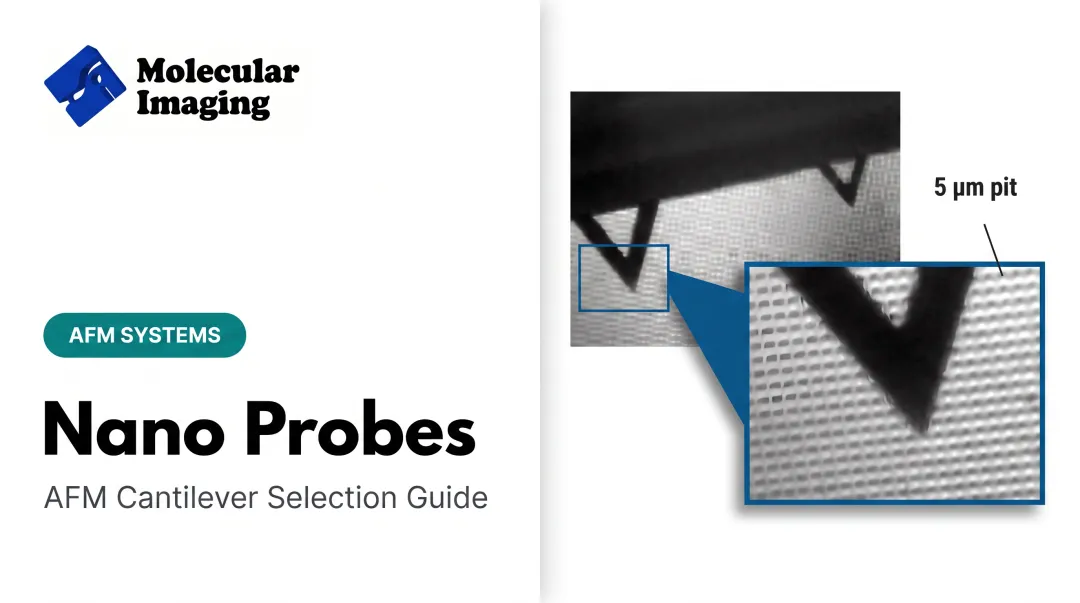
Switch fields entirely, and "silicon probes" means something completely different. In AFM and STM research, silicon probes refer to microfabricated cantilever-and-tip assemblies, made from either silicon or silicon nitride, used to physically scan a sample's surface and generate nanoscale topographic maps.
Specs That Actually Matter Here
Unlike neural probes, there's no impedance or electrode coating to think about. Instead, three mechanical properties determine imaging performance:
- Spring constant — how stiff the cantilever is, affecting how much force it applies to the sample
- Resonance frequency — how fast the cantilever oscillates, relevant for tapping and dynamic imaging modes
- Tip radius of curvature — how sharp the tip is, which directly controls lateral resolution
These three factors need to match the AFM operating mode you're running and the sample you're imaging. A stiff, sharp tip built for tapping mode on a hard material behaves very differently than a soft, flexible cantilever suited to contact mode on a delicate polymer.
Where These Probes Get Used
AFM silicon and silicon nitride probes show up across a surprisingly broad range of research:
- Battery material characterization — tracking solid-electrolyte interphase formation on lithium-ion electrodes in situ
- Polymer surface analysis — mapping stiffness and morphology changes across blended materials
- Reactive gas and humidity studies — comparing tip adhesion behavior under controlled atmospheric conditions
- Biological imaging in liquid — high-speed imaging of proteins and cells in near-native aqueous environments
Molecular Imaging's web store carries a working set of these consumables:
- Contact mode silicon cantilevers (ContSi-10)
- Silicon nitride contact cantilevers (S3N4-10)
- Liquid tapping cantilevers in silicon (Si-L-10) and silicon nitride (Si3N4-L-10) for biological and polymer work in aqueous media
The store also stocks calibration samples, including a 500 nm grid standard and gold-on-mica substrates. Environmental control accessories cover temperature-controlled stages rated from -30°C to 250°C and isolation chambers for humidity, gas, and low-oxygen work.

Protecting Your Investment
A few habits go a long way toward extending probe life:
- Handle cantilevers with anti-static tools and clean tweezers only
- Store probes in their original packaging away from humidity and dust
- Recalibrate spring constant and sensitivity after any suspected tip contact or crash
- Avoid scanning abrasive samples with tips intended for soft-sample imaging
Choosing the Right Silicon Probe for Your Research
The right silicon probe depends on your sample, imaging mode, and how much tip–sample force you can tolerate.
Silicon vs. Silicon Nitride Cantilevers
- Silicon cantilevers — stiffer shanks and sharper tips; a common pick for tapping mode on harder materials
- Silicon nitride cantilevers — more flexible and durable; often favored for contact mode, fluid imaging, and soft or biological samples
Match spring constant and tip radius to the surface. A stiff silicon lever on a soft film can damage the sample; an overly soft lever on a rigid surface can blur topography and slow feedback.
Matching Probe to Research Goal
Before you order, pin down the outcome you need:
- Hard materials and thin films — higher-stiffness silicon probes in tapping or non-contact mode
- Soft matter and bio samples — lower-stiffness silicon nitride levers, often in liquid or contact mode
- Electrical or magnetic contrast — conductive, diamond-coated, or coated specialty tips matched to MFM, EFM, or KFM
- Repeatable metrology — consistent tip geometry plus routine calibration standards
If catalog options fall short, Molecular Imaging can help match cantilevers and custom AFM configurations to your mode, environment, and sample type.
Frequently Asked Questions
What are silicon probes?
Silicon probes are microfabricated devices built on a silicon substrate. They support high-density neural recording or nanoscale surface imaging in AFM and STM systems.
What is the electrode potential of silicon?
Silicon itself is a semiconductor substrate, not the active electrode material. Conductive coatings like titanium nitride or PEDOT determine the actual electrochemical and recording properties at each site.
What are Neuropixels?
Neuropixels are a fully integrated silicon probe design capable of recording from hundreds of neurons simultaneously. A single probe packs 384 channels across 960 densely spaced, programmable recording sites.
How do silicon probes differ from wire electrodes?
Silicon probes offer fixed, repeatable electrode geometry, higher channel counts, and more stable performance over time. Wire electrodes are flexible and cheaper but vary in spacing and wear with repeated use.
Can silicon probes be reused across multiple experiments?
Yes. Both neural and AFM silicon probes are generally reusable with proper cleaning and recalibration. Their fabricated, rigid structure resists the gradual wear common in wire electrodes.
What is the difference between silicon and silicon nitride probes in AFM?
Silicon probes are typically stiffer with sharper tips, suited to tapping mode on harder samples. Silicon nitride probes are more flexible and durable, often chosen for contact mode or softer, biological samples.