Picking the wrong mode isn't a minor inconvenience. It can chew through an expensive tip in minutes, distort your topography data, or generate artifacts that look like real features but aren't. Mode selection deserves the same attention researchers give to sample prep, not an afterthought decided by habit.
This guide walks through the three foundational AFM modes, the specialized techniques built on top of them, and a practical framework for matching mode to sample and research goal.
Key Takeaways
- Contact, tapping, and non-contact modes cover most topographic imaging and differ in whether—and how—the tip touches the surface.
- Specialized modes like KPFM, MFM, and EC-AFM extend AFM into electrical, magnetic, and electrochemical property mapping.
- Scan speed and tip-wear settings directly affect data quality; faster isn't always safer.
- Mode choice should follow sample hardness, environment, and the property under study, not convention.
- Modular AFM/STM platforms let one instrument support multiple modes instead of requiring separate dedicated systems.
What Is AFM and Why Do Its Modes Matter?
Atomic force microscopy uses a sharp cantilever tip, often just a few nanometers across, to sense forces between the probe and a sample surface. As the tip scans, those forces become three-dimensional maps of topography—and, depending on the mode, material properties as well.
"Mode" describes two things at once: how the tip is driven — held in constant contact, oscillated near resonance, or hovered above the surface — and which signal drives the feedback loop. That signal might be cantilever deflection, oscillation amplitude, resonance frequency shift, tunneling current, or phase lag.
Why this matters in practice:
- Soft biological tissue can be torn apart by lateral drag forces that a hard semiconductor wafer would never notice.
- Magnetic thin films need a probe sensitive to stray fields, not just topography.
- Ambient humidity introduces capillary forces that behave completely differently than the same experiment run in liquid or vacuum.
Mismatched mode selection is also a documented source of tip degradation and image artifacts. A 2024 tapping-mode study found that raising the scan line frequency from 0.2 Hz to 1 Hz increased estimated tip-diameter growth from 5.8 nm to 20 nm under the tested conditions, and measured surface roughness dropped by as much as 33% at the higher scan speed.

That drop is a clear sign the tip was blunting mid-scan. Settings that seem like minor conveniences can ruin a dataset.
That risk is one reason modular AFM/STM systems have become popular in multi-user labs. Platforms like the 5500 PicoPlus AFM+STM use interchangeable nose cones, letting a researcher switch between contact, tapping, non-contact, and several specialized electrical modes on one instrument rather than owning separate dedicated systems for each technique.
Core Imaging Modes of Atomic Force Microscopy
Nearly every specialized AFM technique in use today builds on three foundational modes. What separates them is whether the tip contacts the surface continuously, intermittently, or not at all, and which signal the feedback loop tracks.
Contact Mode (Static Mode)
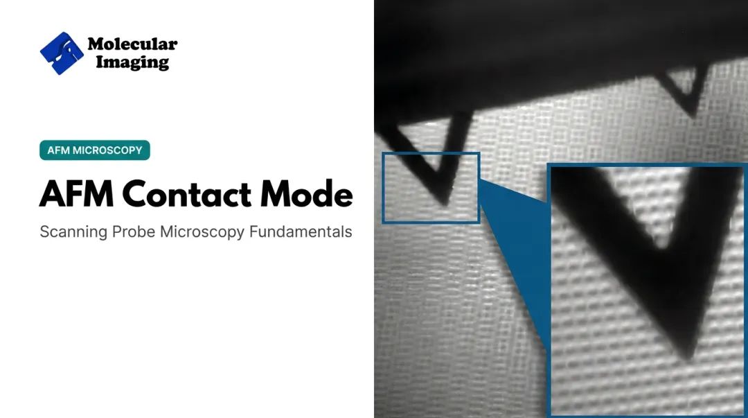
In contact mode, the tip drags continuously across the surface while the system holds cantilever deflection constant, following Hooke's law (F = -kx). As topography changes, the feedback loop adjusts probe height to keep the applied force steady.
Unlike the other two core modes, there's no oscillation involved here. Force is measured directly and continuously, not inferred from amplitude or frequency changes.
Best suited for:
- Hard, rigid, relatively flat samples: crystals, many polymers, semiconductor surfaces
- Lateral and friction force mapping, where continuous contact is actually required
Strengths:
- Fastest of the three core modes
- Simpler, less expensive electronics
Trade-offs:
- Strong lateral shear and capillary forces in ambient air can distort images
- Risk of damage to soft samples and faster tip wear
- Rarely used for biological imaging for these reasons
For labs running contact mode regularly, Molecular Imaging's catalog includes cantilevers built for this purpose: a silicon nitride option (S3N4-10) valued for flexibility on delicate-but-rigid samples, and a silicon variant (ContSi-10) suited to harder substrates.
Tapping Mode (Dynamic / Amplitude Modulation Mode)
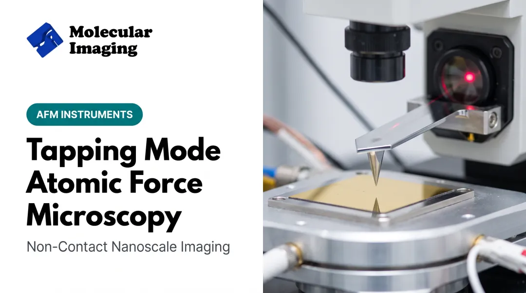
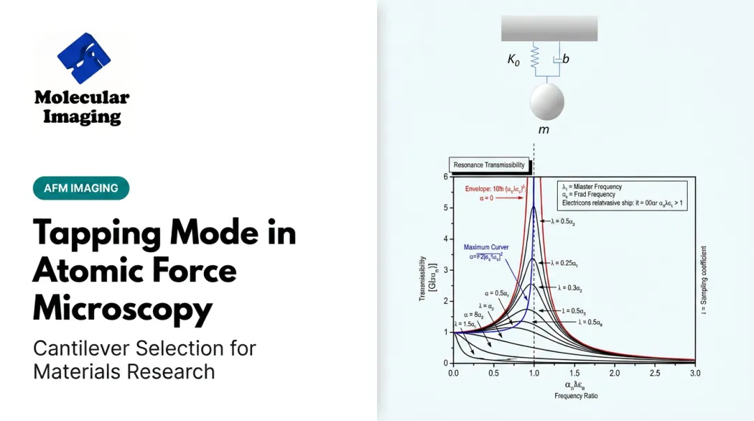
Tapping mode drives the cantilever near its resonance frequency so the tip briefly touches the surface at the bottom of each oscillation cycle. Changes in oscillation amplitude, caused by tip-sample interaction, drive the feedback loop instead of direct deflection.
The intermittent contact virtually eliminates the lateral shear forces that plague contact mode. As a bonus, tapping mode also produces phase-contrast data that captures differences in stiffness and adhesion. That contrast is largely qualitative unless you calibrate carefully.
Best suited for:
- Soft or delicate samples: biological membranes, polymers, thin films
- Imaging in air or liquid, where lateral drag would otherwise cause damage
Tapping-mode AFM has resolved fine biological detail few would have expected from a mechanical probe, including Y-shaped domains and hinge regions on individual antibodies. That resolution depends on matching amplitude and cantilever stiffness to the sample. There is no single universal "right" amplitude setting, despite what some vendor literature implies.
Molecular Imaging offers a tiered set of tapping cantilevers for this reason: the NCH-30-10 for high-frequency, low-damage imaging in air; the NCL-30-10 as a general-purpose middle ground; and the low-frequency FM-10 for the most fragile biological specimens. For liquid work, the Si-L-10 silicon cantilever is designed for aqueous and physiological conditions.
Liquid imaging deserves its own note. Standard acoustic tapping in fluid often introduces resonance artifacts from the surrounding liquid itself. Molecular Imaging's 5500 ILM PicoPlus BioAFM addresses this with MAC Mode, which uses a magnetic field to drive a paramagnetically coated cantilever directly instead of exciting it acoustically.
Cleaner oscillation and gentler intermittent contact make MAC Mode practical for native-state aqueous samples such as:
- Chromatin
- Plasmid DNA
- Protein S-layer assemblies
Non-Contact Mode (Frequency Modulation Mode)
Non-contact mode keeps the cantilever oscillating slightly above its resonance frequency without the tip ever touching the sample. Long-range van der Waals forces shift that resonance frequency, and the feedback loop tracks the shift to maintain a constant tip-sample distance.
This keeps the probe entirely in the attractive-force regime, never dipping into repulsive contact the way tapping mode briefly does.
Best suited for:
- Ultra-soft samples that can't tolerate any contact
- True atomic-resolution imaging, historically demonstrated in ultra-high vacuum on metals, semiconductors, and insulators
UHV is no longer an absolute requirement. Researchers have achieved atomic-resolution frequency-modulation imaging in water, Tris buffer, and even cell-culture medium using stiff sensors. That work extends non-contact imaging into biologically relevant liquids once thought out of reach for this technique.
Trade-offs:
- In ambient air, an adsorbed fluid layer often overwhelms the weak non-contact signal
- Slowest of the three core modes, which limits high-throughput work

Specialized and Advanced AFM Modes
Once you move beyond topography, specialized modes (often built on a tapping or contact-mode foundation) let AFM probe electrical, magnetic, mechanical, and chemical properties directly.
| Mode | What it measures | Typical use case |
|---|---|---|
| Conductive AFM (C-AFM) | Local current or I-V response via a conductive probe | Diagnosing contact failures in semiconductor devices |
| Kelvin Probe Force Microscopy (KPFM) | Surface potential / contact potential difference | Mapping p-, n-, and n+-doped silicon regions |
| Piezoresponse Force Microscopy (PFM) | Electromechanical response to an AC bias | Visualizing ferroelectric domains in thin films |
| Magnetic Force Microscopy (MFM) | Magnetostatic stray fields via a magnetic probe | Mapping magnetic domain patterns in alloys |
| Force Spectroscopy | Force versus tip-sample extension | Single-molecule unfolding studies |
| Electrochemical AFM (EC-AFM) | Electrode topography/mechanics under potential control | In-situ battery and corrosion research |
Force Spectroscopy
Force spectroscopy pulls detailed mechanical data from a single molecule. Early landmark work stretching individual titin proteins recorded a sawtooth force pattern with roughly 25–28 nm periodicity, unfolding individual immunoglobulin domains at forces between 150 and 300 pN depending on pulling speed.
That resolution needs the right liquid cell and cantilever pair. Molecular Imaging's liquid-tapping cantilevers (Si-L-10, Si3N4-L-10) and biomolecule immobilization protocols are built for this class of experiment.
Electrical Modes: C-AFM, KPFM, and PFM
On the electrical side, C-AFM setups use a conductive contact cantilever for current and resistance sensing alongside topography.
KPFM and PFM similarly rely on conductive probes like the EFM-10 cantilever plus lock-in electronics, often through a MAC III controller. Together they extract surface potential or piezoelectric response from the raw deflection signal.
Electrochemical AFM (EC-AFM)
EC-AFM is widely used in battery, corrosion, and photovoltaic research because it lets researchers watch an electrode surface change in real time as potential is applied.
Molecular Imaging's 5500 Series supports this with liquid cells rated for acids, bases, and organic solvents, three-electrode electrochemical configurations, and nanoelectrode integration for combined AFM-SECM measurements. Typical targets include SEI layer formation on battery anodes and corrosion pit growth.

How to Choose the Right AFM Mode for Your Research
Mode selection should follow the sample and the question you're asking, not whichever technique happens to be most familiar. Five factors drive the decision in practice.
Sample hardness and fragility. Soft biological or polymer samples generally call for tapping or non-contact mode. Hard, rigid materials (crystals, metals, most semiconductors) can tolerate contact mode without issue.
Imaging environment. Ambient air, liquid, or vacuum conditions physically narrow your options. True frequency-modulation non-contact imaging has traditionally needed UHV, though liquid-phase atomic resolution is now achievable with the right sensor.
Property of interest. Topography alone is one question; electrical, magnetic, mechanical, or chemical mapping is another entirely. Each specialized mode requires a matched cantilever — conductive coatings for C-AFM and PFM, magnetic coatings for MFM.
Throughput needs. Production and quality-control environments usually favor faster contact or tapping approaches when speed matters more than exhaustive property mapping.
Instrument flexibility. Buying a dedicated system per mode adds up fast in cost and lab space. A platform like the 5500 PicoPlus AFM+STM supports AFM, STM, tapping mode, MAC Mode, C-AFM, KPFM, EFM, and MFM through a modular nose cone—eight modes on one instrument without a full system swap.

Environmental accessories matter too. Temperature-controlled stages spanning -30°C to 250°C and isolation chambers for humidity, gas composition, and low-oxygen conditions extend which modes are viable for a given experiment.
That control is especially important for battery research and polymer phase-transition studies, where atmosphere is not optional.
Conclusion
AFM's real value comes from its range, not any single technique. Contact, tapping, and non-contact modes handle the overwhelming majority of topographic imaging needs, while specialized modes open the door to electrical, magnetic, mechanical, and electrochemical insight that topography alone can't provide.
Matching sample properties, environment, and research goals to the right mode, instead of defaulting to whatever is familiar, produces clean, reliable data and protects tips from unnecessary wear.
Labs evaluating new, replacement, or repaired AFM/STM systems do not have to guess their way through mode selection. Molecular Imaging builds modular platforms and pairs them with direct technical support, so you can add modes as research needs evolve rather than locking into one technique for the life of the instrument.
Frequently Asked Questions
What are the three main modes of atomic force microscopy?
Contact, tapping (dynamic), and non-contact are the three foundational AFM modes. They differ in whether and how the tip touches the sample and which signal (deflection, amplitude, or frequency shift) drives the feedback loop.
What is the difference between contact mode and tapping mode AFM?
Contact mode drags the tip continuously across the surface, while tapping mode oscillates the tip and briefly touches down once per cycle. That intermittent contact in tapping mode greatly reduces lateral shear forces on the sample.
Which AFM mode is best for imaging biological samples?
Tapping mode is the standard choice for soft biological samples because of its gentle, intermittent contact. Techniques like MAC Mode extend this further for imaging in liquid, where conventional acoustic tapping can introduce noise artifacts.
Can a single AFM system perform multiple imaging modes?
Yes. Modular AFM/STM platforms with interchangeable nose cones, like the 5500 PicoPlus AFM+STM, let researchers switch between contact, tapping, non-contact, and specialized electrical or electrochemical modes on one instrument.
What is non-contact AFM mode used for?
Non-contact mode suits ultra-soft samples and true atomic-resolution imaging, typically in ultra-high vacuum. Researchers have also demonstrated it in liquid environments using stiff sensors.
How do I know which AFM mode is right for my sample?
Consider sample hardness, the imaging environment (air, liquid, or vacuum), and the specific property you need to measure. When it's unclear, consulting an AFM instrumentation specialist can save both time and tips.