That matters because many biological and electrochemical samples simply aren't themselves once you dry them out or pull them into a vacuum chamber. Proteins denature. Membranes collapse. Battery electrodes stop behaving like battery electrodes. Researchers studying these systems need to see them wet, hydrated, and functioning.
This article covers what AFM actually is, why liquid imaging changes the game for biology and materials science, how the instrumentation works, where it's applied, and what to look for when choosing a system built for liquid-phase work.
Key Takeaways
- AFM reaches nanometer-to-sub-nanometer resolution on fully immersed samples, which other techniques do not achieve in liquid
- Liquid imaging keeps biological and electrochemical samples in their native hydrated state
- Open or closed liquid cells with tapping or FM-AFM modes enable reliable liquid-phase imaging
- Choose systems for cantilever sensitivity, environmental control, and cross-environment modularity
What Is Atomic Force Microscopy?
AFM is a form of scanning probe microscopy (SPM). Instead of using light or electrons to form an image, it "feels" a surface with a sharp tip mounted on a flexible cantilever. Gerd Binnig, Calvin Quate, and Christoph Gerber introduced the technique in 1986, giving scientists a practical way to map surfaces at the nanometer scale in air or liquid.
AFM does three things well:
- Topographic imaging — mapping surface height with sub-nanometer precision
- Force measurement — quantifying interactions between the tip and sample, down to individual molecular bonds
- Surface manipulation — moving, cutting, or rearranging material at the nanoscale (nanolithography, nanografting)
Three Foundational Imaging Modes
Three primary modes cover most AFM work:
- Contact mode — the tip drags across the surface, maintaining constant force
- Tapping mode — the cantilever oscillates, tapping the surface intermittently
- Non-contact mode — the tip hovers just above the surface, sensing attractive forces

How Expensive Is an AFM System?
Pricing swings widely with capability. Industry vendor data puts general laboratory-grade systems in the $20,000 to $300,000 range, with entry-level or educational units typically below $30,000 and advanced research platforms above $100,000, according to AFM Workshop's published price list.
Modularity, automation, and accessories such as liquid cells and environmental stages all push cost higher.
Molecular Imaging's lineup spans the same range: the Benchtop 5400 is a more accessible entry point, while the 5500 PicoPlus AFM+STM and 5500 ILM PicoPlus BioAFM sit at the research-grade end. Configuration drives cost heavily, so an accurate figure usually means a direct quote rather than a published sheet alone.
AFM's practical edge over electron microscopy is simpler: it does not need a vacuum or a conductive sample coating. That is what makes liquid imaging possible—something electron microscopes cannot offer.
Why AFM in Liquid Matters for Research
Biological structure and function depend on hydration. Strip the water away and a protein folds differently, a membrane loses its bilayer arrangement, a living cell stops being alive. Researchers studying these systems in their natural state need a technique that doesn't force a trade-off between resolution and reality.
An AFM liquid sample is a specimen submerged in a liquid medium (buffer, electrolyte, or aqueous solution) and held in a specialized liquid cell during scanning. The tip operates directly in that fluid rather than in air.
Liquid vs. Air and Vacuum Imaging
Imaging in ambient air introduces capillary and meniscus forces. A thin water layer forms around the tip and sample, distorting force measurements and dragging on the probe. Liquid imaging removes those problems:
- Eliminates the air-water interface that drives capillary forces
- Reduces tip drag and force-measurement distortion
- Sidesteps surface contamination common in air and vacuum imaging
The resolution payoff is real. Researchers using frequency-modulation AFM (FM-AFM) resolved lateral hydration periodicities of 0.53 nm and 0.31 nm on a mineral surface submerged in aqueous potassium chloride solution, capturing a full three-dimensional map in just 1.5 minutes, according to research published in Nature Communications. That's atomic-scale detail, acquired while the sample stayed wet the entire time.
Force sensitivity in liquid AFM reaches down to a few piconewtons, in the same range as optical tweezers, a gold standard for single-molecule work. A widely cited Nature Methods comparison lists AFM's force-spectroscopy range at 10 to 10,000 piconewtons, against optical tweezers' 0.1 to 100 piconewtons, per the journal's methods review.
That overlap makes single-bond and single-molecule studies possible that dried or vacuum-based methods can't touch.
How Liquid AFM Works: Modes and Liquid Cells
Getting clean data in liquid isn't automatic. It requires the right mode, the right cell, and hardware tuned for a fundamentally different physical environment than air.
Imaging Modes Used in Liquid
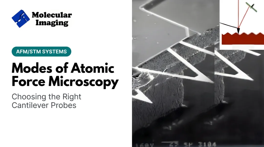
Tapping mode (dynamic mode) dominates liquid imaging because it minimizes the shear forces that damage soft samples. Contact mode drags the tip continuously across the surface — fine for rigid materials, destructive for a lipid membrane or a loosely bound protein. Intermittent tapping reduces that lateral stress considerably.
Molecular Imaging's platforms take this a step further with MAC Mode (Magnetic AC Mode), which drives the cantilever magnetically instead of acoustically. That avoids the spurious resonances and instability that acoustic excitation can introduce in a liquid cell — a known headache for anyone imaging soft biological material in fluid.
For researchers chasing atomic or submolecular resolution, FM-AFM is the tool. It requires low-noise deflection sensors and stiffer cantilevers than typical biological tapping-mode probes, since softer cantilevers are more susceptible to thermal (Brownian) noise in liquid. This combination is what enabled the sub-nanometer hydration mapping and single-DNA-molecule resolution referenced earlier.
Liquid Cell Types
Two broad categories cover most experimental needs:
- Open (microcell) liquid cells — simple design, minimal setup, fast learning curve. Good for quick dunk-and-scan work where you don't need liquid exchange mid-experiment.
- Closed (environmental) liquid cells — sealed systems that support liquid flow-through and inert gas atmospheres. Better suited for electrochemical experiments or time-course studies, though they take longer to master.

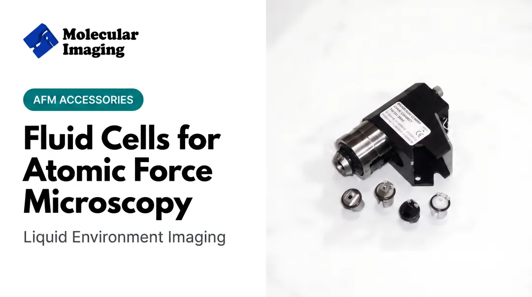
Molecular Imaging offers a wide range of liquid cell designs across its 5500 PicoPlus and 5420 PicoAFM platforms, including configurations compatible with acids, bases, and organic solvents — useful when electrochemistry work calls for aggressive fluids rather than plain buffer.
Instrumentation Considerations
Liquid imaging changes cantilever behavior. Viscous damping from the surrounding fluid reduces the cantilever's quality factor (Q) substantially compared to air, meaning probes selected for air-based tapping mode often perform poorly once submerged. Liquid-specific cantilevers, like the silicon and silicon nitride liquid tapping probes in Molecular Imaging's catalog, are built to maintain stable oscillation under that added hydrodynamic drag.
Environmental control matters too. Molecular Imaging's temperature-controlled sample stages cover -30°C to 250°C, supporting phase-transition and thermal studies that need stability well beyond room temperature. Pair that with an environmental isolation chamber, and researchers can control humidity, gas composition, and oxygen levels alongside temperature, which is essential for reactive or air-sensitive liquid experiments.
Applications and Challenges of AFM in Liquid
Liquid AFM is essential wherever samples must stay native, hydrated, or electrochemically active.
Life sciences lean on it heavily:
- DNA and protein imaging in near-physiological buffer
- Lipid bilayer structure down to individual headgroups
- Living cell surfaces, membrane dynamics, and mechanics in real time
Materials science and electrochemistry use it just as extensively:
- In-situ battery electrode studies tracking solid-electrolyte interphase growth as voltage changes
- Corrosion and passive-film analysis on metals under controlled electrochemical potential
- Electrochemical AFM (EC-AFM) correlating surface topography with current or redox activity in real time
| Application Area | What Liquid AFM Reveals |
|---|---|
| DNA/protein imaging | Molecular structure in physiological buffer |
| Lipid bilayers | Individual headgroup arrangement |
| Living cells | Real-time membrane dynamics |
| Battery electrodes | SEI growth under applied voltage |
| Corrosion studies | Passive film thickness and roughness changes |
| Molecular electronics | Simultaneous structure and conductivity mapping |
Molecular electronics research also benefits, where conductive-probe liquid AFM/STM allows simultaneous imaging and electrical measurement of molecular junctions, a combination that is difficult to achieve any other way.
The Practical Challenges
Liquid imaging comes with trade-offs:
- Signal drift from liquid volume changes, temperature shifts, and evaporation over long sessions
- Reduced cantilever sensitivity due to viscous damping and a lower effective Q factor than in air
- Weaker signal-to-noise that often requires specialized low-noise electronics
Purpose-built hardware helps offset these issues. Molecular Imaging's pendulum-style multipurpose scanner uses a lightweight, rigid mechanical loop to limit the drift and vibration sensitivity that liquid imaging amplifies.
Choosing the Right AFM System for Liquid Imaging
Not every AFM handles liquid well. When evaluating a system for fluid-phase work, focus on a short list of features that matter most:
- Liquid cell compatibility — open and closed cell designs, plus tolerance for acids or organic solvents when needed
- Low-noise detection electronics — preserves resolution when viscous damping rises
- Modularity — one platform that switches between air, liquid, and environmental modes
Molecular Imaging's 5500 ILM PicoPlus BioAFM and the broader 5500 PicoPlus Series are built around that flexibility. Interchangeable nose cones support AFM, STM, conductive AFM, and related modes on one scanner head.
Academic advisor Dr. Stuart Lindsay, who co-founded the original Molecular Imaging Corporation in the mid-1990s, brings decades of scanning probe research from Arizona State University into that modular design approach.
Flexibility at purchase only helps if the system stays reliable in daily liquid work. Molecular Imaging backs its liquid AFM platforms with video tutorials, downloadable manuals on electrochemistry and liquid cell setup, and ZoomAFM remote repair support when performance issues arise.

Frequently Asked Questions
What is atomic force microscopy?
AFM is a high-resolution scanning probe technique that images surfaces by measuring the force between a cantilever-mounted tip and the sample. It works in air, vacuum, or liquid, unlike most other microscopy methods.
What is an AFM liquid sample?
It's a specimen submerged in a liquid medium (buffer, electrolyte, or aqueous solution) and held in a specialized liquid cell during scanning. This keeps biological and electrochemical samples in their native, functional state.
How expensive is atomic force microscopy?
Costs vary widely, generally from around $20,000 for basic systems to over $300,000 for advanced research platforms, depending on modularity and accessories. Contact a specialized provider like Molecular Imaging for an accurate quote based on your configuration.
What liquids can be used for AFM imaging?
Common choices include water and physiological buffers; electrochemistry work often needs a specific electrolyte instead. Match the liquid to the sample so the surface stays stable during the scan.
Can AFM image living cells?
Yes. AFM images live cells directly in buffer or culture medium, enabling real-time studies of membrane dynamics and cell mechanics without fixing or drying the sample.
What's the difference between open and closed liquid cells?
Open cells are simpler and quicker for basic dunk-and-scan imaging. Closed cells are sealed, support liquid flow-through, and allow controlled atmospheres. They suit electrochemical or time-course experiments better, though they need more setup expertise.